组成图示
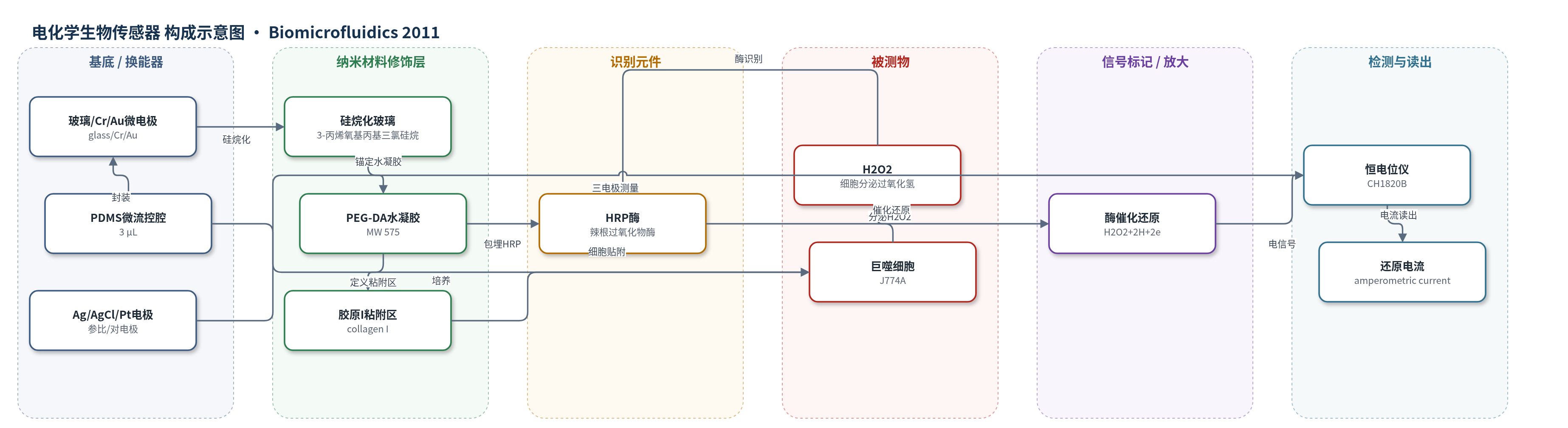
点击图片查看大图 · 依据论文自动绘制
传感器类型
电化学生物传感器
检测对象
过氧化氢(hydrogen peroxide, H2O2);样品基质:巨噬细胞(J774A)微流控培养液/细胞分泌上清(DMEM)
检测原理
巨噬细胞在 PMA 刺激下产生氧化应激并分泌 H2O2。H2O2 扩散至电极上方 PEG-DA 水凝胶中的 HRP 活性位点,HRP 催化 H2O2 还原为水(H2O2 + 2H+ + 2e- → H2O),酶自身被氧化。工作电极固定在 -0.2 V vs Ag/AgCl,通过直接电子转移将电子传递给氧化态 HRP,使其恢复活性,同时产生与 H2O2 浓度成正比的还原电流。由于 HRP 催化循环,信号较无酶电极增强约 60 倍。微流控腔将细胞限制在电极旁,小体积(3 μL)和静置测量有利于富集分泌物,实现实时安培监测。
检测灵敏度
LOD: 2 μM(原文结果部分记为 ~2 μM);线性范围: 2 μM–500 μM;R^2 = 0.96;灵敏度: 27 μA cm^-2 mM^-1;原文另称线性范围可扩展至 2 mM
效应效果
传感器在 -0.2 V vs Ag/AgCl 下响应时间约 10 s(90% 信号)。含 HRP 电极比无 HRP 凝胶电极灵敏约 60 倍。PMA 刺激约 1000 个 J774A 巨噬细胞后,最大净还原电流变化 8 pA,约 60 min 达峰,随后 60 min 减弱;H2O2 分泌量为 10.3 ± 1.53 μM(n=3)。无 PMA、无细胞加 PMA 及无 HRP 电极对照均无显著还原电流,表明信号来自激活巨噬细胞分泌的 H2O2。作者认为电化学方案比 Amplex Red/HRP 荧光法更稳健、灵敏和可重复,H2O2 产生速率 0.03 nmol/10^3 cells/h 与文献 0.05–0.13 nmol/10^3 cells/h 相当,可用于临床免疫细胞氧化爆发监测。
传感器的构成
- 基底/换能器电极:玻璃载玻片溅射 15 nm Cr 粘附层和 100 nm Au 层,光刻湿法刻蚀成 300 μm 直径 Au 微电极,作为工作电极。
- 硅烷锚定层:3-丙烯氧基丙基三氯硅烷修饰玻璃,用于共价锚定 PEG 水凝胶。
- 酶载水凝胶修饰层:聚乙二醇二丙烯酸酯(PEG-DA, MW 575)预聚体加入光引发剂 2-羟基-2-甲基丙苯酮、辣根过氧化物酶(HRP)和 2% 戊二醛,UV 光刻成 600 μm 水凝胶圆盘,固定 HRP 并防止电极污损。
- 识别/催化元件:HRP 包埋于 PEG-DA 水凝胶中,催化 H2O2 还原并实现直接电子转移。
- 细胞粘附区:第二层无 HRP 的 PEG 水凝胶光刻形成 300 μm 宽环形裸露玻璃区,孵育 I 型胶原(collagen I)促进巨噬细胞贴附。
- 微流控封装层:聚二甲基硅氧烷(PDMS)双通道微流控腔(6×10×0.1 mm,体积 3 μL),封装电极并控制细胞与刺激物输送。
- 辅助电极:流通式 Ag/AgCl 参比电极置于出口,Pt 丝对电极置于入口,与 Au 工作电极组成三电极系统。
中文摘要
本文报道了一种用于芯片上检测免疫细胞氧化应激的电化学生物传感器。作者将微型金电极与巨噬细胞无缝集成,用于检测细胞分泌的过氧化氢(H2O2)。采用聚乙二醇(PEG)水凝胶光刻技术,一方面将辣根过氧化物酶(HRP)固定在电极表面,另一方面在电极附近定义细胞贴附区域。微图案化电极被封装于聚二甲基硅氧烷(PDMS)微流控通道中,并与流通式 Ag/AgCl 参比电极和 Pt 丝对电极组成三电极电化学池。巨噬细胞仅贴附于裸露玻璃区域,而不粘附于非污损 PEG 水凝胶表面。该微器件对 H2O2 的灵敏度为 27 μA cm^-2 mM^-1,检出限为 2 μM。器件可实现巨噬细胞在电极附近的可控接种、细胞激活以及 H2O2 释放的实时监测。未来该平台可用于监测巨噬细胞对病原体的反应或研究微图案化细胞培养中的炎症信号。
英文摘要
Seamless integration of biological components with electrochemical sensors is critical in the development of microdevices for cell analysis. The present paper describes the integration miniature Au electrodes next to immune cells (macrophages) in order to detect cell-secreted hydrogen peroxide (H(2)O(2)). Photopatterning of poly(ethylene glycol) (PEG) hydrogels was used to both immobilize horseradish peroxidase molecules onto electrodes and to define regions for cell attachment in the vicinity of sensing electrodes. Electrodes micropatterned in such a manner were enclosed inside poly(dimethylsiloxane) fluid conduits and incubated with macrophages. The cells attached onto the exposed glass regions in the vicinity of the electrodes and nowhere else on the non-fouling PEG hydrogel surface. A microfluidic device was converted into an electrochemical cell by placing flow-through Ag∕AgCl reference and Pt wire counter electrodes at the outlet and inlet, respectively. This microdevice with integrated H(2)O(2)-sensing electrodes had sensitivity of 27 μA∕cm(2) mM with a limit of detection of 2 μM. Importantly, this microdevice allowed controllable seeding of macrophages next to electrodes, activation of these cells and on-chip monitoring of H(2)O(2) release in real time. In the future, this biosensor platform may be utilized for monitoring of macrophage responses to pathogens or for the study of inflammatory signaling in micropatterned cell cultures.