组成图示
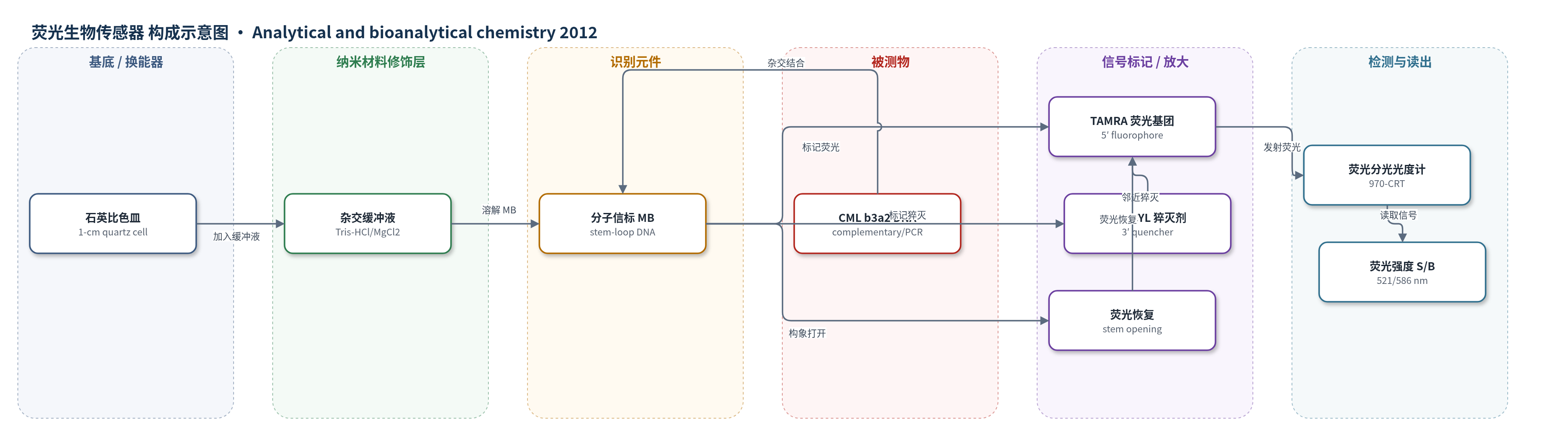
点击图片查看大图 · 依据论文自动绘制
传感器类型
荧光生物传感器
检测对象
慢性髓系白血病(CML)b3a2 型 BCR/ABL 融合基因 DNA 片段(CML b3a2 DNA fragment)、PCR 扩增产物(PCR amplification products);样品基质为静脉血来源 cDNA/PCR 产物及 K562 细胞 cDNA
检测原理
MB 为茎环结构,5′端 TAMRA 与 3′端 DABCYL 在茎区邻近,通过荧光共振能量转移(FRET)猝灭 TAMRA 荧光,背景低。当 CML b3a2 互补 DNA 存在时,MB 环区与靶标杂交,茎环打开,TAMRA 与 DABCYL 距离增大,猝灭效率下降,TAMRA 荧光恢复。荧光强度或信背比(S/B)随互补 DNA 浓度增加而升高,在 4.0×10⁻⁹–3.2×10⁻⁸ mol/L 线性。单碱基错配或非互补序列不能有效打开茎环,荧光变化小,从而实现选择性检测。该方法未使用酶促或链式放大,主要依赖 MB 内禀构象变化实现信号转导。
检测灵敏度
LOD: 6.0×10⁻¹⁰ mol/L (S/N=3);线性范围: 4.0×10⁻⁹–3.2×10⁻⁸ mol/L;相关系数: 0.9973;回归方程: Y(S/B)=4.0354X(C)+3.1754
效应效果
该方法对互补序列、单碱基错配序列和非互补序列表现出明显选择性,单碱基错配时荧光信号显著低于互补序列,非互补序列几乎无荧光恢复。真实样品检测中,MB 空白平均荧光强度为 3.578,RSD 为 2.1%;阳性 PCR 扩增样品平均荧光强度为 16.344,RSD 为 7.2%;阴性样品平均荧光强度为 5.674,RSD 为 6.3%,阳性与阴性差异明显。荧光检测结果与琼脂糖凝胶电泳结果一致,可区分阳性真实样品、阴性真实样品和 K562 阳性细胞。作者认为该方法比染色体分析、Southern blot、FISH 和常规 PCR 凝胶电泳更快速、简便、灵敏,且避免溴化乙锭污染,可用于 CML 早期诊断和预后监测。
传感器的构成
- 检测容器:1-cm 石英比色皿(quartz cell),溶液相荧光检测容器
- 识别元件:分子信标(MB),序列 5′-TAMRA-CGCTGC-AGA GTT CAA AAG CCC TTC-GCAGCG-DABCYL-3′,环区识别 CML b3a2 互补 DNA
- 信号标记物:TAMRA(tetramethoxyl rhodamine),5′端荧光基团,杂交后荧光恢复
- 猝灭剂:DABCYL(4-(2-methyl-on-amino-azobenzene) benzoate),3′端猝灭剂,无靶标时猝灭 TAMRA
- 杂交缓冲液:Tris–HCl/MgCl2 缓冲液(20 mM Tris–HCl、10 mM MgCl2,pH 7.5),维持 MB 发夹与杂交
- 信号读出:970-CRT 荧光分光光度计(λex=521 nm,λem=586 nm),检测荧光强度变化
中文摘要
本文建立了一种基于分子信标的荧光生物传感器方法,用于检测慢性髓系白血病(CML)相关基因片段及 PCR 扩增产物。作者设计了一种分子信标(MB),其环区序列依据 CML b3a2 型 BCR/ABL 融合基因 DNA 序列设计,5′端标记荧光基团四甲基罗丹明(TAMRA),3′端标记猝灭剂 DABCYL。未杂交时,MB 呈发夹结构,TAMRA 与 DABCYL 空间邻近,荧光被猝灭;当 MB 与互补 DNA 杂交后,茎环结构打开,猝灭剂远离荧光基团,荧光强度恢复并增强。在优化条件下,荧光信号与互补 DNA 浓度在 4.0×10⁻⁹–3.2×10⁻⁸ mol/L 范围内呈线性关系,相关系数为 0.9973,检出限为 6.0×10⁻¹⁰ mol/L(S/N=3)。该方法具有较高选择性,可区分单碱基错配序列,并成功用于检测 278 bp 的 CML 短链 DNA 片段及真实样品 PCR 产物,有望用于 CML 临床诊断与预后监测。
英文摘要
A novel fluorescence method has been established for the determination of gene fragment and PCR amplification products related to chronic myelogenous leukemia (CML). A molecular beacon (MB) which comprises a DNA loop section, a pair of fluorophore (tetramethoxyl rhodamine, TAMRA), and a quencher (4-(2-methyl-on-amino-azobenzene) benzoate, DABCYL) was designed. The loop sequence of MB was designed according to the DNA sequence relating to CML (type b3a2) which contained a single-stranded oligonucleotide. Before hybridization, the fluorescence from the TAMRA had been quenched by the DABCYL. After hybridization with the complementary DNA, the quencher will become far away from the TAMRA, and the fluorescence intensity detected will increase. Changes in the fluorescence intensity have a linear relationship with the concentration of complementary DNA (C) in the range of 4.0 × 10(-9)-3.2 × 10(-8) mol/L, with a correlation coefficient of 0.9973; the detection limit was 6.0 × 10(-10) mol/L (S/N = 3). The developed method has high selectivity, which can be used to discriminate single-base mismatch sequence. The method has been applied to detect the short-stranded CML DNA fragment (278 bp) with high sensitivity. This approach is a promising method for the detection of CML in real samples for medical diagnostics.