组成图示
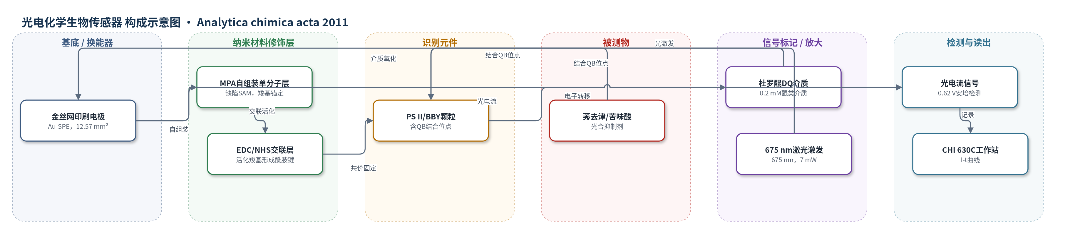
点击图片查看大图 · 依据论文自动绘制
传感器类型
光电化学生物传感器
检测对象
莠去津(atrazine)、苦味酸(picric acid);样品基质:MES 测量缓冲液(含 0.2 mM DQ),面向环境水样分析
检测原理
传感器以 PS II 为识别元件。光照下 PS II 反应中心发生电荷分离,电子沿电子传递链到达 QB 位点;在体外,杜罗醌(DQ)作为类质体醌类似物结合/接近 QB 位点并接受电子,被还原的 DQ 通过缺陷 MPA SAM 和 BBY 膜沟槽扩散至金电极,在 0.62 V 下氧化,形成可测光电流。当莠去津或苦味酸等抑制剂存在时,它们竞争结合 QB 位点,阻止 DQ 接受电子,使光电流随抑制剂浓度增加而下降。信号变化通过恒电位安培法记录,无需额外标记或酶放大。
检测灵敏度
LOD: 1.15 nM(atrazine)、157 nM(picric acid,表2为157.5 nM);动态响应范围: 1 nM–1 μM(atrazine);IC50: 49 nM(atrazine)、784 nM(picric acid);Hill slope: 0.82(atrazine)、1.13(picric acid);R^2 = 0.9984(atrazine)、0.9936(picric acid)
效应效果
BBY 颗粒传感器在 12.5 mm² 电极面积下光电流为 20–35 nA,明显优于 PS II 核心颗粒的 1.00±0.75 nA;与 BSA–戊二醛凝胶固定相比,SAM 固定响应更快、信噪比更好。传感器 24 h 内光电流约减半,前 2 h 基本稳定;10 nM 莠去津重复测量变异系数约 5%(n=3),洗涤后再生几乎 100%。莠去津 LOD 1.15 nM 低于欧盟 MRL 50 μg/L(232 nM),接近饮用水 MRL 0.1 μg/L(2.32 nM);苦味酸 LOD 157 nM 高于 BSA 凝胶法 25 nM,但优于荧光淬灭法 2 μM。作者认为其适合非选择性早期预警,但尚不能区分不同抑制剂。
传感器的构成
- 基底/换能器电极:金丝网印刷电极(Au-SPE),含金工作电极(12.57 mm²)、金对电极和银伪参比电极,陶瓷基底,用于光电化学换能。
- 自组装单分子层:巯基丙酸(MPA)SAM,2 mM 在 75/25% 乙醇/水中形成,故意保留针孔缺陷,提供羧基锚定并允许介质传输。
- 交联活化层:EDC/NHS 处理 MPA 羧基,形成氨基反应性中间体,与光合材料胺基形成酰胺键。
- 识别元件:菠菜 PS II 核心颗粒或 BBY 颗粒(PS II 富集膜碎片),含 QB 结合位点,用于结合光合抑制剂。
- 信号介质:杜罗醌(DQ,0.2 mM),作为类质体醌类似物在 QB 位点接受电子,还原后扩散至电极氧化产生光电流。
- 光源与读出:675 nm 激光二极管(7 mW)照射,CHI 630C 电化学工作站,在 0.62 V 恒电位下记录 I–t 光电流。
中文摘要
本文报道了一种基于光合反应中心的光电化学生物传感器。作者利用巯基丙酸(MPA)在金丝网印刷电极(Au-SPE)上形成故意存在缺陷的自组装单分子层(SAM),并通过 EDC/NHS 将菠菜来源的光系统 II(PS II)核心颗粒或 BBY 颗粒共价固定于电极表面。缺陷单分子层和 BBY 颗粒形成的沟槽结构有利于杜罗醌(DQ)介质向电极和 QB 位点传输。在 675 nm 激光照射下,PS II 发生光诱导电荷分离,电子经 QB 位点传递给 DQ,还原 DQ 在金电极上氧化产生光电流。光合抑制剂与 QB 位点结合后阻断电子传递,使光电流下降。该传感器用于检测莠去津和苦味酸,检出限分别为 1.15 nM 和 157 nM,并显示比 BSA–戊二醛凝胶固定方式更快的响应和更好的信噪比。
英文摘要
Photosynthetic reaction centers were immobilized onto gold screen-printed electrodes (Au-SPEs) using a self-assembled monolayer (SAM) of mercaptopropionic acid (MPA) which was deliberately defective in order to achieve effective mediator transfer to the electrodes. The pure Photosystem II (PS II) cores from spinach immobilize onto the electrodes very efficiently but fair badly in terms of photocurrent response (measured using duroquinone as the redox mediator). The cruder preparation of PS II known as BBY particles performs significantly better under the same experimental conditions and shows a photocurrent response of 20-35 nA (depending on preparation) per screen-printed electrode surface (12.5mm(2)). The data was corroborated using AFM, showing that in the case of BBY particles a defective biolayer is indeed formed, with grooves spanning the whole thickness of the layer enhancing the possibility of mass transfer to the electrodes and enabling biosensing. In comparison, the PS II core layer showed ultra-dense organization, with additional formation of aggregates on top of the single protein layer, thus blocking mediator access to the electrodes and/or binding sites. The defective monolayer biosensor with BBY particles was successfully applied for the detection of photosynthesis inhibitors, demonstrating that the inhibitor binding site remained accessible to both the inhibitor and the external redox mediator. Biosensing was demonstrated using picric acid and atrazine. The detection limits were 1.15 nM for atrazine and 157 nM for picric acid.