组成图示
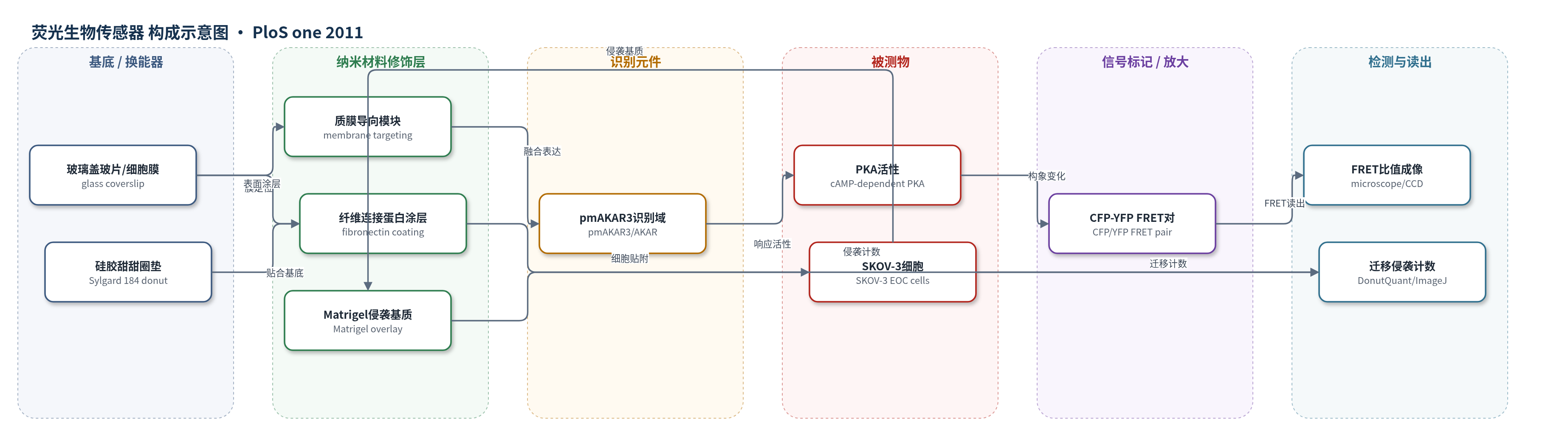
点击图片查看大图 · 依据论文自动绘制
传感器类型
荧光生物传感器
检测对象
蛋白激酶A活性(Protein Kinase A activity, PKA activity);样品基质:SKOV-3 卵巢癌细胞(SKOV-3 EOC cells,细胞内/质膜区),Leibovitz L-15 培养基,纤维连接蛋白涂层玻璃盖玻片,Matrigel 三维基质
检测原理
pmAKAR3 是质膜导向的 FRET 型 PKA 活性报告蛋白,由 CFP、AKAR 响应域和 YFP 组成。当细胞内 cAMP 依赖性 PKA 活性升高时,AKAR 域发生构象变化,改变 CFP 与 YFP 之间的距离或取向,从而提高 FRET 效率;显微镜分别采集 CFP、YFP 和 FRET 通道,经背景扣除后计算 YFP/CFP 或 FRET 比值,比值升高表示 PKA 活性增强。由于传感器被导向质膜,可分辨迁移或侵袭细胞前缘的局部 PKA 激活。该检测为比值型自参考荧光读出,不依赖外源电子供体或酶放大;迁移/侵袭行为则通过硅胶甜甜圈垫限制单层、Matrigel 覆盖和 ImageJ 计数读出。
检测灵敏度
原文未报告 LOD、线性范围、灵敏度斜率或相关系数。
效应效果
pmAKAR3 在 SKOV-3 细胞中显示 PKA 特异性:共表达 mCherry-PKI 后前缘和整体 PKA 活性几乎消除。FRET 成像显示迁移细胞前缘 PKA 活性升高;>80% 表达 pmAKAR3 的迁移细胞显示前缘活性(LE/Cyto FRET ratio >1.2),共表达 sAKAPis 后仅 <30%,RIAD 或对照仍 >80%。侵袭实验中 >80% 侵袭细胞显示侵袭前缘 PKA 活性,sAKAPis 后仅 <10%。donut assay 使用 <6000 细胞,较划痕实验更好保留 ECM 和细胞活力;GM6001 抑制 Matrigel 侵袭,验证侵袭。作者主张 PKA 活性和 AKAP 锚定是 EOC 迁移、侵袭和转移的关键机制。
传感器的构成
- 基底/换能器:玻璃盖玻片(glass coverslips)与 SKOV-3 细胞膜,承载 pmAKAR3 并支撑 FRET 成像
- 膜定位层:质膜导向模块(plasma membrane-directed targeting),使 pmAKAR3 定位于细胞膜
- 识别元件:pmAKAR3 的 AKAR 响应域(AKAR),响应 PKA 催化活性并改变构象
- 信号标记:CFP 与 YFP 荧光蛋白对(CFP/YFP),作为 FRET 供体/受体,PKA 活性改变 FRET 比值
- 读出层:倒置荧光显微镜(Nikon Eclipse TE-2000E)、60x/1.4 NA Plan Apo 油镜、冷却 CCD 相机,采集 CFP/YFP/FRET 图像
- 样品基质:SKOV-3 卵巢癌细胞(SKOV-3 EOC cells),Leibovitz L-15 培养基,10 ng/ml EGF 刺激迁移
- 迁移/侵袭检测结构:Sylgard 184 硅胶甜甜圈垫(donut gasket)、纤维连接蛋白(fibronectin)涂层和 Matrigel 基质,用于限制细胞单层并测量迁移/侵袭
中文摘要
上皮性卵巢癌(EOC)是致死率最高的妇科恶性肿瘤,部分原因是其临床隐匿性转移。理解 EOC 扩散和侵袭机制可能提供抗转移治疗新靶点或转移病检测新方法。cAMP 依赖性蛋白激酶(PKA)在 EOC 中常失调,且 PKA 活性和通过 A 激酶锚定蛋白(AKAPs)的亚细胞定位是细胞骨架动力学和细胞迁移的重要调节因子。因此,我们研究 PKA 和 AKAP 功能在 EOC 细胞迁移和侵袭中的作用。使用质膜导向 PKA 生物传感器 pmAKAR3 和改良迁移/侵袭实验,我们证明 PKA 在迁移 SKOV-3 EOC 细胞的前缘被激活,抑制 PKA 活性阻断 SKOV-3 细胞迁移。此外,虽然这些细胞前缘内 PKA 活性由 II 型调节亚基(RII)锚定介导,但抑制 RI 或 RII PKA 亚基锚定均阻断细胞迁移。重要的是,我们首次证明在 SKOV-3 细胞侵袭三维细胞外基质过程中,PKA 活性在侵袭前缘上调,并且与迁移类似,抑制 PKA 活性或 AKAP 介导的 PKA 锚定阻断基质侵袭。这些数据首次证明癌细胞侵袭细胞外基质会引发侵袭前缘内 PKA 激活,且 PKA 活性和锚定均对基质侵袭必不可少。这些观察提示 PKA 和 AKAP 活性在 EOC 转移中的作用。
英文摘要
Epithelial ovarian cancer (EOC) is the deadliest of the gynecological malignancies, due in part to its clinically occult metastasis. Therefore, understanding the mechanisms governing EOC dissemination and invasion may provide new targets for antimetastatic therapies or new methods for detection of metastatic disease. The cAMP-dependent protein kinase (PKA) is often dysregulated in EOC. Furthermore, PKA activity and subcellular localization by A-kinase anchoring proteins (AKAPs) are important regulators of cytoskeletal dynamics and cell migration. Thus, we sought to study the role of PKA and AKAP function in both EOC cell migration and invasion. Using the plasma membrane-directed PKA biosensor, pmAKAR3, and an improved migration/invasion assay, we show that PKA is activated at the leading edge of migrating SKOV-3 EOC cells, and that inhibition of PKA activity blocks SKOV-3 cell migration. Furthermore, we show that while the PKA activity within the leading edge of these cells is mediated by anchoring of type-II regulatory PKA subunits (RII), inhibition of anchoring of either RI or RII PKA subunits blocks cell migration. Importantly, we also show--for the first time--that PKA activity is up-regulated at the leading edge of SKOV-3 cells during invasion of a three-dimensional extracellular matrix and, as seen for migration, inhibition of either PKA activity or AKAP-mediated PKA anchoring blocks matrix invasion. These data are the first to demonstrate that the invasion of extracellular matrix by cancer cells elicits activation of PKA within the invasive leading edge and that both PKA activity and anchoring are required for matrix invasion. These observations suggest a role for PKA and AKAP activity in EOC metastasis.