组成图示
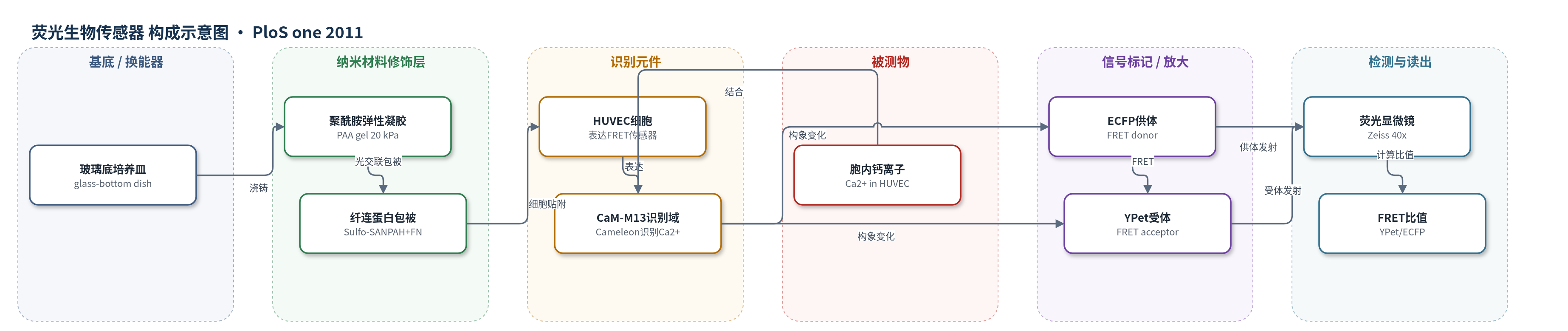
点击图片查看大图 · 依据论文自动绘制
传感器类型
荧光生物传感器
检测对象
细胞内钙离子(intracellular calcium, Ca2+);样品基质:活人脐静脉内皮细胞(HUVEC)胞质/细胞内环境,培养于聚酰胺弹性凝胶上。
检测原理
机械振动使聚酰胺凝胶局部变形,HUVEC 膜牵张激活通道开放,胞外 Ca2+ 内流;局部钙升高激活磷脂酶 C(PLC),产生三磷酸肌醇(IP3),IP3 受体介导内质网(ER)钙释放,形成全局胞内钙升高。基因编码 FRET 钙传感器中,Ca2+ 与钙调蛋白-M13(CaM-M13)结合后改变 ECFP 与 YPet 的距离/取向,使 FRET 效率变化。用 420/20 nm 激发 ECFP,分别采集 475/40 nm 供体与 535/25 nm 受体发射,计算 YPet/ECFP 比值;比值随胞内 Ca2+ 浓度升高而增大,从而实时反映钙信号时空分布。
检测灵敏度
—
效应效果
装置在凝胶中产生约140 Hz、振幅约70 μm的探针振动,细胞位置最大位移约13–14 μm、最大应变约30%–40%。对照刺激下80%细胞呈全局钙响应、20%呈局部响应。EGTA螯合胞外钙后多数响应消失;Gd3+(p=0.007)和链霉素(p=0.02)阻断牵张激活通道后全局响应显著减少;thapsigargin(p=1×10⁻⁵)和U73122(p=7×10⁻⁴)使响应转为局部;ML-7(p=3×10⁻⁴)和cytochalasin D(p=0.01)抑制肌动球蛋白收缩/肌动蛋白完整性后全局响应下降。作者认为该非接触振动装置可研究亚细胞力学转导。
传感器的构成
- 基底/换能器:玻璃底培养皿(glass-bottom dish)与聚酰胺弹性凝胶(PAA gel,8% acrylamide/0.13% bis,约20 kPa),承载细胞并传递局部机械振动。
- 表面修饰:Sulfo-SANPAH 光交联后包被牛纤连蛋白(bovine fibronectin, FN),促进 HUVEC 贴附。
- 识别元件:基因编码改良 Cameleon 中的钙调蛋白(calmodulin, CaM)与 M13 肽,结合胞内 Ca2+ 引起构象变化。
- 信号标记物:增强青色荧光蛋白(ECFP)作为 FRET 供体,YPet 作为 FRET 受体,Ca2+ 结合改变 YPet/ECFP 能量转移效率。
- 表达/递送系统:腺病毒载体(Adeno-X)携带 FRET 生物传感器质粒感染 HUVEC,实现胞内表达。
- 读出模块:Zeiss 荧光显微镜,420/20 nm 激发 ECFP,475/40 nm 与 535/25 nm 分别采集 ECFP 与 YPet 发射,计算 FRET 比值。
中文摘要
本研究设计了一种新装置,可在柔性弹性凝胶上产生局部机械振动,从而在亚细胞位置对人脐静脉内皮细胞(HUVEC)进行机械刺激。采用基于荧光共振能量转移(FRET)的钙生物传感器(改良型 Cameleon,以 ECFP 和 YPet 为供体/受体)监测机械刺激下细胞内钙浓度的时空分布。结果显示,多数细胞在机械刺激后出现覆盖整个细胞体的胞内钙浓度升高(全局响应)。用 EGTA 螯合胞外钙,或用链霉素、氯化镓阻断质膜牵张激活钙通道,可显著抑制机械刺激引起的钙响应。内质网钙泵抑制剂 thapsigargin 或磷脂酶 C 抑制剂 U73122 处理则主要使钙响应局限于刺激位点附近。破坏肌动蛋白丝(cytochalasin D)或抑制肌动球蛋白收缩(ML-7)也抑制全局钙响应。因此,HUVEC 的全局钙响应依赖于质膜牵张激活通道介导的钙内流,随后经磷脂酶 C 激活产生三磷酸肌醇(IP3),触发内质网钙释放。该机械刺激装置还可作为研究细胞在亚细胞水平感知力学线索分子机制的有力工具。
英文摘要
A new device was designed to generate a localized mechanical vibration of flexible gels where human umbilical vein endothelial cells (HUVECs) were cultured to mechanically stimulate these cells at subcellular locations. A Fluorescence Resonance Energy Transfer (FRET)-based calcium biosensor (an improved Cameleon) was used to monitor the spatiotemporal distribution of intracellular calcium concentrations in the cells upon this mechanical stimulation. A clear increase in intracellular calcium concentrations over the whole cell body (global) can be observed in the majority of cells under mechanical stimulation. The chelation of extracellular calcium with EGTA or the blockage of stretch-activated calcium channels on the plasma membrane with streptomycin or gadolinium chloride significantly inhibited the calcium responses upon mechanical stimulation. Thapsigargin, an endoplasmic reticulum (ER) calcium pump inhibitor, or U73122, a phospholipase C (PLC) inhibitor, resulted in mainly local calcium responses occurring at regions close to the stimulation site. The disruption of actin filaments with cytochalasin D or inhibition of actomyosin contractility with ML-7 also inhibited the global calcium responses. Therefore, the global calcium response in HUVEC depends on the influx of calcium through membrane stretch-activated channels, followed by the release of inositol trisphosphate (IP3) via PLC activation to trigger the ER calcium release. Our newly developed mechanical stimulation device can also provide a powerful tool for the study of molecular mechanism by which cells perceive the mechanical cues at subcellular levels.