组成图示
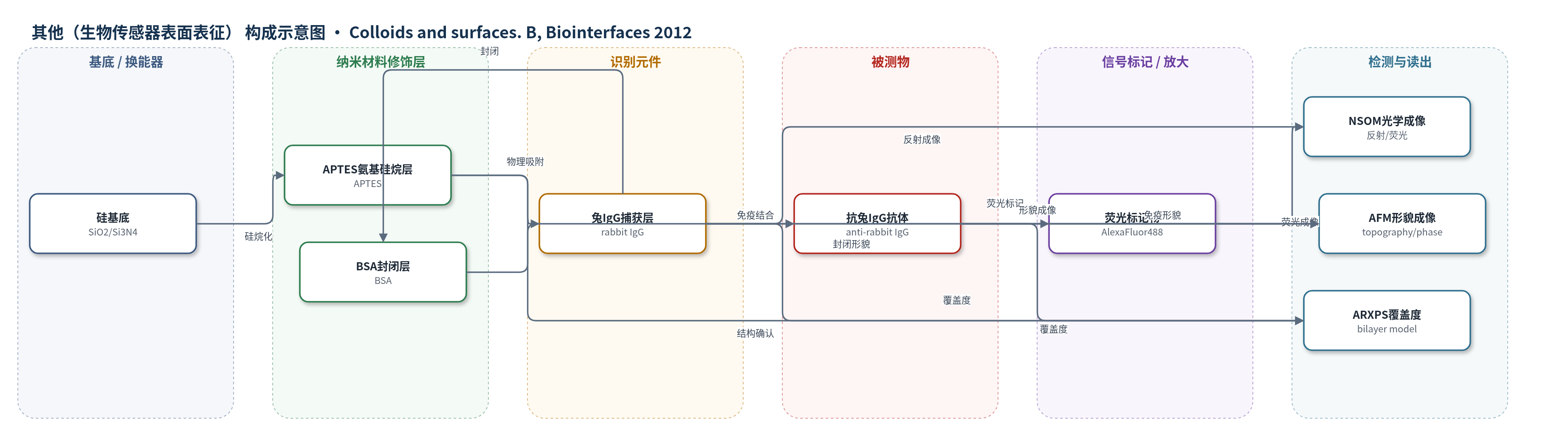
点击图片查看大图 · 依据论文自动绘制
传感器类型
其他(生物传感器表面表征)
检测对象
抗兔IgG抗体(anti-rabbit IgG antibody);样品基质:0.05 M磷酸盐缓冲液(pH 7.4,免疫反应含10 mg/mL BSA)
检测原理
该模型表面通过APTES在SiO2/Si3N4上形成氨基界面,兔IgG以物理吸附方式固定,BSA封闭剩余位点,抗兔IgG抗体随后与固定兔IgG发生特异性免疫结合。免疫结合使表面形成第二蛋白层和免疫复合物,导致蛋白覆盖度、表面粗糙度及横向结构尺度增加;若抗体携带AlexaFluor488,则结合量增加使荧光强度增强。ARXPS利用不同光电子出射角下N 1s与Si 2p光电子强度的角度依赖,通过双层衰减模型定量蛋白层厚度;AFM和NSOM分别提供横向纳米结构、均一性和光学信号分布。该过程无酶催化或核酸放大,主要依赖免疫识别和荧光标记/光电子能谱读出。
检测灵敏度
原文未报告LOD、线性范围、灵敏度斜率或相关系数。
效应效果
NSOM显示蛋白层均匀:反射强度172±81 kHz和228±50 kHz,RMS粗糙度约0.3 nm和0.7 nm;荧光强度612±178 Hz。ARXPS给出Si3N4兔IgG覆盖度1.9±0.1 mg/m2,封闭后1.5±0.1,免疫后4.0±0.4;SiO2为1.1±0.1、1.2±0.3和3.4±0.4 mg/m2。直接吸附anti-rabbit IgG覆盖度为1.2±0.1(Si3N4)和0.7±0.1(SiO2)mg/m2。免疫反应使覆盖度平均增加约270%(结论中约280%),AFM特征尺寸2whm增加约190%,间距1/k增加约165%;封闭在误差内不显著改变覆盖度和横向尺度,但可能伴随IgG与BSA部分交换。作者认为该表征有助于优化硅基生物传感器。
传感器的构成
- 基底:硅片,SiO2(约1000 nm热生长二氧化硅)或Si3N4(约150 nm氮化硅),作为硅基换能器基底
- 硅烷化修饰层:3-氨基丙基三乙氧基硅烷(APTES),水解缩合形成氨基有机硅烷薄膜,提供氨基界面
- 识别元件:兔γ-球蛋白/免疫球蛋白G(rabbit IgG),物理吸附形成捕获蛋白层
- 封闭剂:牛血清白蛋白(BSA,10 mg/mL),封闭自由结合位点
- 检测抗体/信号标记:抗兔IgG抗体(anti-rabbit IgG antibody),部分用AlexaFluor488荧光标记,用于免疫结合与荧光成像
- 参考表面:直接吸附BSA或anti-rabbit IgG的APTES硅烷化基底,用于对照
中文摘要
在硅基生物传感器中,氨基有机硅烷修饰表面上生物识别蛋白层的组成与结构决定器件的灵敏度和特异性。本研究采用多种光谱与显微方法,系统表征了在SiO2或Si3N4基底上构建的模型生物传感器表面:先用3-氨基丙基三乙氧基硅烷(APTES)修饰,再物理吸附兔γ-球蛋白(IgG),用牛血清白蛋白(BSA)封闭,并特异性结合抗兔IgG抗体;同时制备直接吸附BSA或抗兔IgG抗体的硅烷化基底作为参考。X射线光电子能谱(XPS)证实了蛋白/氨基有机硅烷/硅多层结构;近场扫描光学显微镜(NSOM)在反射和荧光模式下验证了蛋白覆盖的均匀性;角度分辨XPS(ARXPS)结合双层模型定量蛋白表面覆盖度;原子力显微镜(AFM)分析蛋白层内部横向结构;高分辨ARXPS揭示碳功能团的垂直取向。结果表明,封闭步骤在实验误差内不增加表面覆盖度和横向结构尺度,而免疫反应后覆盖度与各项结构尺度显著上升;所有蛋白层中极性碳功能团均优先朝向硅烷化硅基底,且该取向不受覆盖度、封闭和特异性结合影响。
英文摘要
Composition and structure of biorecognition protein layers created on silicon substrates modified with amino-organosilanes determine the sensitivity and specificity of silicon based biosensing devices. In the present work, diverse spectroscopic and microscopic methods were applied to characterize model biosensor surfaces, formed on Si(3)N(4) or SiO(2) by modification with (3-aminopropyl)triethoxysilane, coating with rabbit gamma-globulins (IgGs) through physical adsorption, blocking with bovine serum albumin (BSA) and specific binding of an anti-rabbit IgG antibody. In addition, silanized substrates with directly adsorbed BSA or anti-rabbit IgG antibody were examined as reference surfaces. The protein/amino-organosilane/silicon structure of all surfaces was confirmed by X-ray photoelectron spectroscopy. Homogeneity of protein coverage was verified with near-field scanning optical microscope, working in reflection and fluorescence mode. Surface coverage with proteins was determined with angle-resolved XPS using a previously established bilayer approach. Inner structure of protein layers was examined with atomic force microscopy. Vertical arrangement of carbon functional groups was revealed by high resolution ARXPS. Combined spectroscopic and microscopic data reveal the complex character of interactions with the immobilized IgG molecules during blocking with BSA and immunoreaction with anti-IgG antibody. Within experimental error, neither surface coverage nor lateral structural scales of protein layer (provided by Fourier and auto-correlation analysis of topographic and phase images) increase during blocking procedure. On the other hand, coverage and all structural measures rise considerably after immunoreaction. In addition, it was found that polar functional groups orient towards substrate for all protein layers, independently of coverage, prior to and after both blocking and specific binding.