组成图示
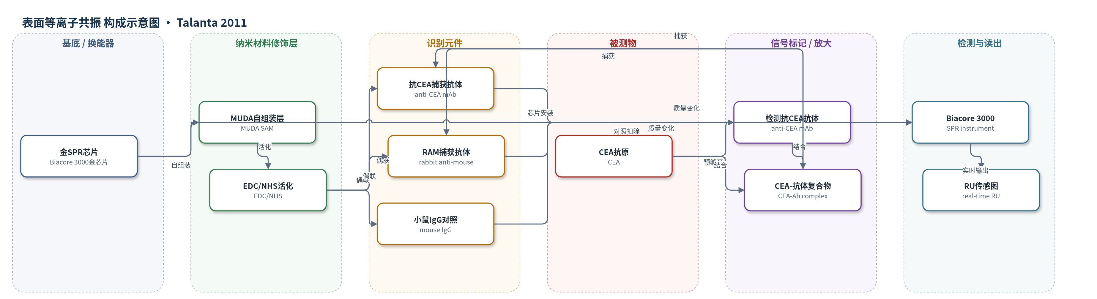
点击图片查看大图 · 依据论文自动绘制
传感器类型
表面等离子共振(SPR)生物传感器
检测对象
癌胚抗原(carcinoembryonic antigen, CEA);样品基质为含5 μg/mL BSA的PBS缓冲液(文中未实际检测血清)
检测原理
在裸金SPR芯片上先形成MUDA自组装单分子层,经EDC/NHS活化后共价固定捕获抗体。检测时,CEA抗原先与检测用抗CEA抗体在含BSA的PBS中预孵育,形成抗原-抗体复合物;复合物注入芯片后,在夹心法中与表面抗CEA抗体结合,或在RAM-capture法中被兔抗鼠IgG捕获。结合事件使金表面附近质量/折射率增加,导致SPR共振角或响应单位(RU)变化,Biacore 3000实时记录传感图。信号经对照通道扣除后随CEA浓度升高而增大;预孵育和夹心/捕获格式通过增加结合质量实现无标记信号放大。
检测灵敏度
LOD: 3 ng ml−1;线性范围: 3 ng ml−1–400 ng ml−1;R² = 1.00(夹心法)、0.99(RAM-capture);斜率: 6.065 RU/(ng ml−1)(夹心法)、1.87 RU/(ng ml−1)(RAM-capture)
效应效果
选择性方面,CEA对小鼠IgG对照表面的非特异结合为零,5 μg/mL BSA非特异结合约3±2 RU,检测抗体非特异结合5.1±5.2 RU。夹心法在3–400 ng/mL范围内响应为30–802 RU,RAM-capture为13–430 RU;Rmax由标准捕获法215 RU提高到RAM-capture 428 RU和夹心法734 RU。数据为三次重复平均,作者称重现性良好,但未报告RSD和实际血清加标回收率。与ELISA相比,该SPR方法无标记、实时、步骤简单;相比部分条带/QCM/SPR CEA方法,其3 ng/mL检出限可满足非吸烟者早期筛查需求,作者认为可用于癌症生物标志物分析。
传感器的构成
- 基底/换能器:裸金SPR传感芯片(Biacore 3000 gold sensor chip),提供表面等离子共振换能表面
- 自组装单分子层:11-巯基十二烷酸(MUDA)SAM,经氮气等离子清洗后浸入2 mM MUDA过夜,形成羧基功能化金表面
- 化学偶联层:EDC/NHS活化MUDA羧基形成NHS酯,用于抗体氨基共价偶联
- 捕获识别元件:鼠源抗CEA单克隆抗体(anti-CEA mAb,Abcam)固定于夹心法通道;兔抗鼠IgG(RAM)固定于RAM-capture通道;小鼠IgG固定于对照通道
- 封闭与封端:BSA封闭非特异结合位点,乙醇胺(ethanolamine)封端未反应NHS酯
- 检测识别元件:Sigma抗CEA单克隆抗体(anti-CEA mAb)作为检测抗体,与CEA预孵育后形成抗原-抗体复合物
- 样品基质:含5 μg/mL BSA的PBS缓冲液,用于配制CEA标准品并降低非特异吸附
中文摘要
本研究开发了一种基于表面等离子共振(SPR)的高灵敏免疫传感器,用于检测肿瘤标志物癌胚抗原(CEA)。在裸金SPR传感芯片表面,首先用11-巯基十二烷酸(MUDA)形成自组装单分子层,再固定抗体。作者比较了直接捕获法和夹心法等不同免疫分析格式;为提高信号,将CEA抗原与检测/捕获抗体预先孵育后再注入芯片,并用Biacore 3000仪器实时记录响应。夹心法和兔抗鼠(RAM)捕获法的检出限均为3 ng/mL,动态检测范围为3–400 ng/mL,相关系数分别为1.00和0.99。动力学分析显示,标准捕获法最大响应Rmax为215 RU,RAM捕获法为428 RU,夹心法为734 RU。结果表明,基于夹心格式的SPR免疫传感器对CEA检测具有高灵敏度和重现性,是癌症生物标志物分析的一种有前景方法。
英文摘要
An immunoassay in optimised conditions with a highly sensitive surface plasmon resonance (SPR) based biosensor was developed for the detection of the cancer biomarker carcinoembryonic antigen (CEA). Different formats of the immunoassay were initially investigated on the surface of the gold sensor chip. A self-assembled monolayer (SAM) was formed on the gold chip using 11-mercaptoundecanoic acid (MUDA), before the immobilisation of the antibodies was conducted. The assay was then formed in a direct capture and a sandwich assay. In order to increase the sensor signal the CEA antigen was incubated with the detection/capture antibody before it was injected to the sensor chip surface and the results were recorded in real-time using the Biacore 3000 instrument. A detection limit of 3 ng ml(-1) CEA was obtained with a dynamic detection range from 3 ng ml(-1) to 400 ng ml(-1) with correlation coefficients of 1.00 and 0.99 for the sandwich and rabbit anti-mouse (RAM) capture assay. Kinetic data analysis was performed for the standard capture test and subsequently for the developed assays and R(max) showed an increase from 215 RU for the standard capture test to 428 RU for the RAM-capture assay and 734 RU for the sandwich assay, respectively. The developed SPR immunosensor using the sandwich assay format showed high sensitivity and reproducibility for CEA detection which makes it a promising procedure for cancer biomarker analysis.