组成图示
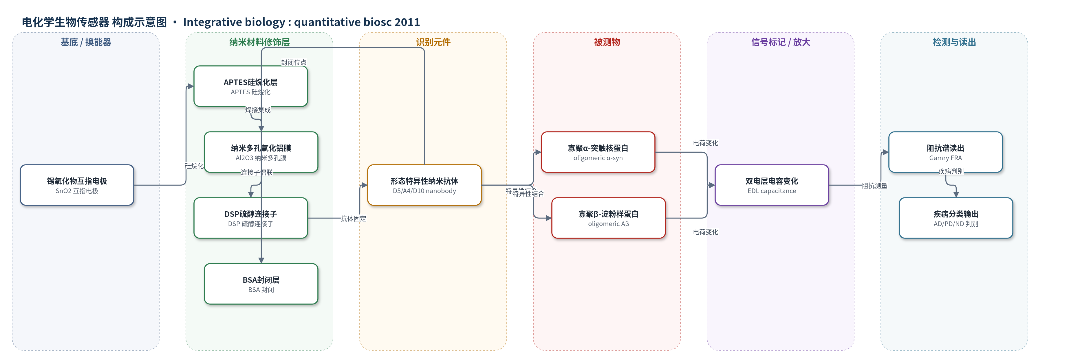
点击图片查看大图 · 依据论文自动绘制
传感器类型
电化学生物传感器
检测对象
寡聚α-突触核蛋白(oligomeric alpha-synuclein, α-syn)、寡聚β-淀粉样蛋白(oligomeric beta-amyloid, Aβ);样品基质:人死后脑脊液(post-mortem human CSF,AD、PD、MSA、认知正常对照)
检测原理
传感器采用无标记电化学阻抗谱(EIS)免疫检测。形态特异性纳米抗体通过 APTES 和 DSP 连接子固定在纳米多孔氧化铝膜内的 SnO2 电极表面,BSA 封闭非特异位点。当 CSF 中的寡聚 α-syn 或 Aβ 与对应纳米抗体结合时,纳米孔固液界面电荷分布发生变化,导致双电层电容改变。由于结合过程不依赖酶促反应或电子供体,界面传导为非法拉第过程,可用 RC 等效电路描述。仪器在 100 Hz、100 mV p-p 正弦激励下测量阻抗,低频下双电层电容主导阻抗谱,阻抗变化随结合量增加而增大,从而反映目标寡聚体浓度。形态特异性纳米抗体提供选择性,纳米孔阵列提高界面信号并限制抗原。
检测灵敏度
LOD: 1 picogram ml−1(α-syn 校准)
效应效果
该传感器在纯品校准中显示对 α-syn 的特异性结合,BSA 对照无信号,无纳米抗体时背景信号显著低于结合信号。在 10 倍稀释的人死后 CSF 中,D5 和 A4 均可检测相应寡聚体。9 例样本(3 例 AD、3 例 PD 样含 2 例 PD 和 1 例 MSA、3 例正常对照)中,A4 可区分 AD 与 PD/ND;D5 在 PD 最高、AD 较低、ND 和 MSA 最低,并能区分 PD 与 MSA。作者用共同参考 AD1 归一化芯片间差异并多次重复,但未报告 RSD、回收率或与 ELISA 的定量对比。作者认为该技术有望用于神经退行性疾病早期诊断。
传感器的构成
- 基底/换能器电极:PCB 平台上的互指锡氧化物(SnO2)工作电极与对电极,提供电化学阻抗谱(EIS)换能
- 纳米多孔膜:250 nm 厚、孔径 200 nm 的纳米多孔氧化铝(Al2O3)膜,焊接于电极上形成纳米孔阵列,电隔离并限制抗原
- 硅烷化修饰层:3-氨基丙基三乙氧基硅烷(APTES)胺功能化电极表面,提供氨基用于后续连接
- 连接子层:3,3'-二硫代双琥珀酰亚胺基丙酸酯(DSP)硫醇连接子,共价偶联纳米抗体
- 识别元件:形态特异性纳米抗体(nanobody/ScFv),D10 结合所有 α-syn 用于校准,D5 识别寡聚 α-syn,A4 识别寡聚 Aβ
- 封闭剂:牛血清白蛋白(BSA)封闭未结合氨基位点,降低非特异结合
- 微流控腔:圆形硅胶腔限制液体并防止蒸发,最大工作体积 1.6 ml
- 信号读出:频率响应分析仪/电化学工作站(Gamry Reference 600 potentiostat)施加 100 mV p-p 正弦电压,测量 100 Hz 阻抗变化
中文摘要
蛋白质错误折叠与聚集是许多严重神经退行性疾病的关键特征,因此表征脑脊液(CSF)中β-淀粉样蛋白(Aβ)和α-突触核蛋白(α-syn)等关键形式与形态的浓度谱,可作为这些疾病的有效诊断方法。CSF 中 tau 和 Aβ 水平已被证明对阿尔茨海默病(AD)具有良好生物标志物前景。然而,许多神经退行性疾病的发病与进展同包括不同 Aβ 和 α-syn 聚集种类在内的可溶性寡聚体密切相关,因此特异性检测并定量 CSF 中每种毒性蛋白种类,可能为无症状诊断和疾病鉴别提供简单而准确的手段。本文表明,利用高选择性形态特异性试剂结合灵敏电子生物传感器,可便捷检测人 CSF 样本中不同蛋白形态。进一步表明,这些形态特异性试剂可区分来自 AD、帕金森病(PD)和认知正常来源的死后 CSF 样本。研究提示,检测特定寡聚聚集种类作为神经退行性疾病敏感生物标志物具有巨大前景。
英文摘要
Protein misfolding and aggregation is a critically important feature in many devastating neurodegenerative diseases, therefore characterization of the CSF concentration profiles of selected key forms and morphologies of proteins involved in these diseases, including β-amyloid (Aβ) and α-synuclein (a-syn), can be an effective diagnostic assay for these diseases. CSF levels of tau and Aβ have been shown to have great promise as biomarkers for Alzheimer's disease. However since the onset and progression of many neurodegenerative diseases have been strongly correlated with the presence of soluble oligomeric aggregates of proteins including various Aβ and a-syn aggregate species, specific detection and quantification of levels of each of these different toxic protein species in CSF may provide a simple and accurate means to presymptomatically diagnose and distinguish between these diseases. Here we show that the presence of different protein morphologies in human CSF samples can be readily detected using highly selective morphology specific reagents in conjunction with a sensitive electronic biosensor. We further show that these morphology specific reagents can readily distinguish between post-mortem CSF samples from AD, PD and cognitively normal sources. These studies suggest that detection of specific oligomeric aggregate species holds great promise as sensitive biomarkers for neurodegenerative disease.