组成图示
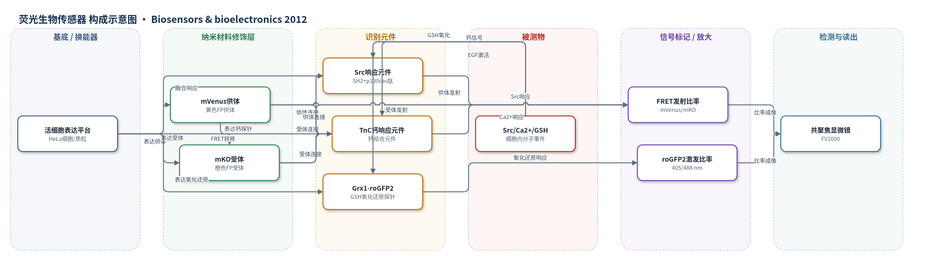
点击图片查看大图 · 依据论文自动绘制
传感器类型
荧光生物传感器
检测对象
Src激酶活性(Src kinase activity)、钙离子(Ca2+)、谷胱甘肽氧化还原电位(GSH redox potential, GSH/GSSG);样品基质:HeLa活细胞
检测原理
YO-Src中,EGF激活Src激酶后磷酸化p130cas底物肽,SH2结构域结合磷酸化肽,使mVenus与mKO的距离或取向改变,FRET效率随之变化,mVenus/mKO发射比率反映Src激酶活性。YO-TnC中,Ca2+结合TnC引起构象变化,改变mKO/mVenus发射比率。Grx1-roGFP2中,GSH/GSSG氧化还原状态改变roGFP2中Cys-Tyr-Cys位点的氧化还原,使405/488 nm激发比率变化。共聚焦显微镜分别采集FRET发射比率和roGFP2激发比率,实现单细胞双参数比率成像;低光谱串扰无需强度校正。
检测灵敏度
未报告LOD、线性范围、灵敏度斜率或相关系数。
效应效果
该组合在HeLa细胞中低串扰:1.4±0.05%的Grx1-roGFP2信号进入mVenus通道,5.2±0.12%的mVenus信号进入roGFP2通道,无需强度校正。控制实验显示H2O2仅改变Grx1-roGFP2的405/488激发比率,不影响YO-Src1.0;EGF仅改变YO-Src1.0发射比率,不影响Grx1-roGFP2。YO-Src对100 ng/ml EGF的ΔR为34.9%±0.7%(n=7),YO-TnC对5 μM ionomycin的ΔR为70.8%±1.8%(n=9),cpmVenus173-TnC-mKO为81.9%±1.1%(n=7);YO-Src1.0动态范围约35%,与ECFP/mCitrine Src传感器(25–35%)相当,Ca2+ YO对(82%)低于优化CFP/YFP对(400%)。YO-Src1.0和YO-TnC1.0可逆。作者主张方法可扩展用于单细胞双分子事件动力学研究。
传感器的构成
- 表达基底:HeLa细胞/质粒载体,承载YO-Src、YO-TnC与Grx1-roGFP2融合蛋白表达
- FRET供体层:mVenus(黄色荧光蛋白,YFP变体)或cpmVenus157/cpmVenus173,作为供体荧光团
- FRET受体层:mKO(橙色荧光蛋白,OFP),作为受体荧光团
- 识别元件:Src响应元件(SH2结构域+p130cas底物肽)或TnC钙响应元件,响应Src磷酸化/Ca2+结合
- 单FP识别层:Grx1-roGFP2(谷胱甘肽还原酶Grx1融合roGFP2),响应GSH/GSSG氧化还原电位
- 信号读出层:共聚焦激光扫描显微镜FV1000,514 nm激发mVenus并收集mVenus/mKO发射,405/488 nm激发roGFP2
中文摘要
基因编码荧光蛋白(FP)生物传感器是无创追踪活细胞分子事件的有力工具。尽管已有多种FP生物传感器,但在单个活细胞中同时成像多个生物传感器(多参数成像)仍是挑战,尚未常规用于解析复杂分子事件网络。本研究建立了一种新型FP生物传感器组合,用于双参数比率荧光成像,由新的荧光共振能量转移(FRET)对mVenus(黄色FP)/mKO(橙色FP)(简称YO)生物传感器和单FP生物传感器Grx1-roGFP2组成。在成像条件下,1.4±0.05%的Grx1-roGFP2信号贡献到mVenus通道,5.2±0.12%的mVenus信号贡献到Grx1-roGFP2通道。作者证明这种低串扰对YO FRET生物传感器和Grx1-roGFP2的比率信号造成可忽略失真。利用该双参数比率成像方法,实现了单个细胞中Src/Ca2+信号与谷胱甘肽(GSH)氧化还原电位的同步成像,这是此前难以实现的。研究进一步提供直接证据表明,表皮生长因子(EGF)诱导的Src信号通过H2O2对GSH氧化还原系统的作用受到负调控,展示了该方法揭示单细胞内不同分子事件新联系的能力。该双参数成像方法具有高度可扩展性。
英文摘要
Genetically coded fluorescent protein (FP)-based biosensors are powerful tools for the non-invasive tracking of molecular events in living cells. Although a variety of FP biosensors are available, the simultaneous imaging of multiple biosensors (multi-parameter imaging) in single living cells remains a challenge and is far from routinely used to elucidate the intricate networks of molecular events. In this study, we established a novel combination of FP biosensors for dual-parameter ratiometric imaging, consisting of a new fluorescence resonance energy transfer (FRET) pair mVenus (yellow FP)/mKOκ (orange FP)-based (abbreviated as YO) biosensor and a single FP-based biosensor Grx1-roGFP2. Under our imaging condition, 1.4±0.05% of Grx1-roGFP2 signal contributes to the mVenus channel and 5.2±0.12% of the mVenus signal contributes to the Grx1-roGFP2 channel. We demonstrate that such low degree of cross-talk causes negligible distortion of the ratiometric signal of the YO-based FRET biosensor and Grx1-roGFP2. By using this dual-parameter ratiometric imaging approach, we achieved simultaneous imaging of Src/Ca(2+) signaling and glutathione (GSH) redox potential in a single cell, which was previously unattainable. Furthermore, we provided direct evidence that epidermal growth factor (EGF)-induced Src signaling was negatively regulated by H(2)O(2) via its effect on GSH-based redox system, demonstrating the power of this dual-parameter imaging approach for elucidating new connections between different molecular events that occur in a single cell. More importantly, the dual-parameter imaging approach described in this study is highly extendable.