组成图示
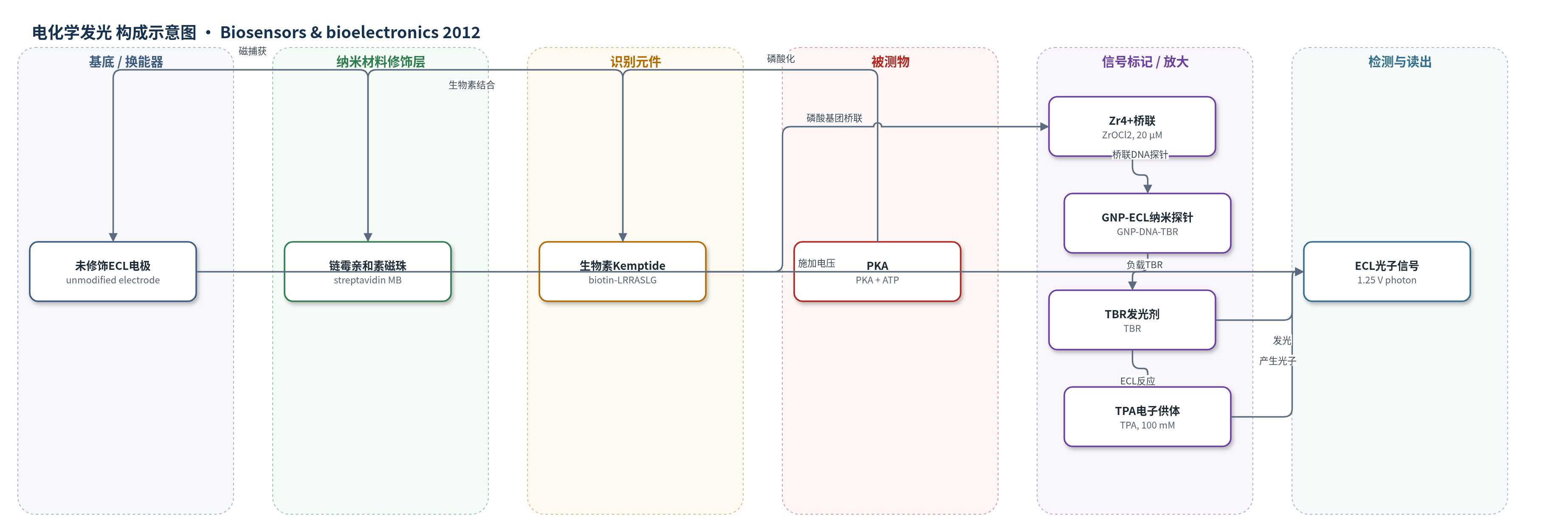
点击图片查看大图 · 依据论文自动绘制
传感器类型
电化学发光(ECL)生物传感器
检测对象
蛋白激酶活性(protein kinase activity,模型蛋白激酶 A,protein kinase A, PKA)、PKA 抑制剂(ellagic acid,鞣花酸);样品基质:含 ATP 的 Tris–HCl/MgCl2 反应缓冲液
检测原理
PKA 在 ATP 存在下催化生物素化 Kemptide 磷酸化,磷酸化产物经生物素–链霉亲和素作用结合到 MB 表面。Zr4+ 与磷酸基团配位,桥联磷酸化肽与 ECL 纳米探针上的磷酸化 DNA 捕获探针,使 GNP 负载的 TBR 被富集至未修饰工作电极附近。施加 1.25 V 后,TBR 与 TPA 发生 ECL 反应并产生光子。PKA 活性越高,磷酸化 Kemptide 越多,捕获的 ECL 纳米探针越多,ECL 强度越大。GNP 负载多个 TBR 实现信号放大,MB 分离去除 ATP、PKA 等未反应组分,降低背景并提高灵敏度。
检测灵敏度
LOD: 0.005 U/mL;线性范围: 0.01 U/mL–50 U/mL;R^2 = 0.9951
效应效果
该传感器对 PKA 的检出限为 0.005 U/mL,线性范围为 0.01–50 U/mL,R^2 = 0.9951,检出限较此前荧光、电化学和比色法低约两个数量级。ECL 纳米探针在 4 ℃ 保存一个月以上仍保持 ECL 活性。对 PKA 抑制剂鞣花酸的 IC50 为 5.33 μM,ECL 强度随抑制剂浓度升高而下降并在 10 μM 附近趋于平台。MB 可去除未反应 ATP 和 PKA 等干扰,简化清洗与电极修饰流程;文中以三次重复误差棒表示重现性,但未给出具体 RSD 数值。作者认为该平台可用于蛋白激酶活性检测、抑制剂筛选及临床诊断与药物研究。
传感器的构成
- 工作电极:未修饰 ECL 工作电极,施加 1.25 V 并检测光子信号
- 磁珠捕获层:链霉亲和素包被磁珠(streptavidin-coated MB,2.8 μm),捕获生物素化磷酸化 Kemptide 并富集至电极表面
- 识别底物:生物素修饰 Kemptide(biotin-LRRASLG),PKA 催化磷酸化后通过生物素–链霉亲和素结合 MB
- 桥联剂:Zr4+(ZrOCl2,20 μM),特异性桥联磷酸化肽与磷酸化 DNA 探针
- ECL 纳米探针:13 nm GNP 负载磷酸化 DNA 捕获探针(5'-HPO4--CCTGAGACGTTTTTTTT-SH-3')和 TBR–cysteamine,提供多价 ECL 信号放大
- 信号标记物:TBR(tris-(2,2'-bipyridyl) ruthenium),ECL 发光剂,与 TPA 反应产生光子信号
- 电子供体:TPA(tripropylamine,100 mM),ECL 反应共反应剂,促进 TBR 发光
中文摘要
本文报道了一种基于磁珠(MB)技术和金纳米颗粒(GNP)信号增强的高灵敏电化学发光(ECL)生物传感器,用于蛋白激酶活性检测及抑制监测。ECL 纳米探针由 GNP 与磷酸化 DNA 捕获探针及三(2,2'-联吡啶)钌(TBR)–半胱胺偶联制备;锆离子(Zr4+)作为特异性桥联剂,介导生物素修饰磷酸化肽与 ECL 纳米探针之间的连接。反应后,链霉亲和素包被 MB 将复合物捕获并富集至未修饰 ECL 电极表面进行发光检测。以蛋白激酶 A(PKA)为模型酶,该传感器检出限达 0.005 U/mL,并可依据 ECL 信号变化定量监测 PKA 抑制剂。MB 分离与未修饰电极检测相结合,使流程简化且可重复,适用于蛋白激酶活性的高灵敏分析。
英文摘要
Herein, we describe a novel electrochemiluminescence (ECL) biosensor for protein kinase activities and inhibition monitoring based on the magnetic beads (MB) technology and signal enhancement of gold nanoparticles (GNP). In this design, ECL nanoprobes were prepared by conjugating GNP with phosphorylated DNA capture probes and tris-(2,2'-bipyridyl) ruthenium (TBR)-cysteamine. Zirconium cations, a specific bridging agent, mediate the linkage between biotin modified phosphorylated peptides and ECL nanoprobes. The complexes were then captured and enriched on the electrode surface by streptavidin-coated MB for ECL reaction. To confirm the feasibility of this biosensor, we employed protein kinase A (PKA) as the model kinase to validate the assay and a satisfactory detection limit of 0.005 U/mL was achieved. The combination of ECL and GNP lays a solid foundation for highly sensitive assay, meanwhile, the coupling of MB surfaces used for separation and capture with unmodified ECL electrode detection results in a greatly simplified and reusable protocol. Thus, our biosensor offers great promise for a highly sensitive and simple assay for protein kinase activity. Furthermore, the inhibition of PKA activity was monitored on the basis of the ECL signals change in response to the concentration of PKA inhibitor.