组成图示
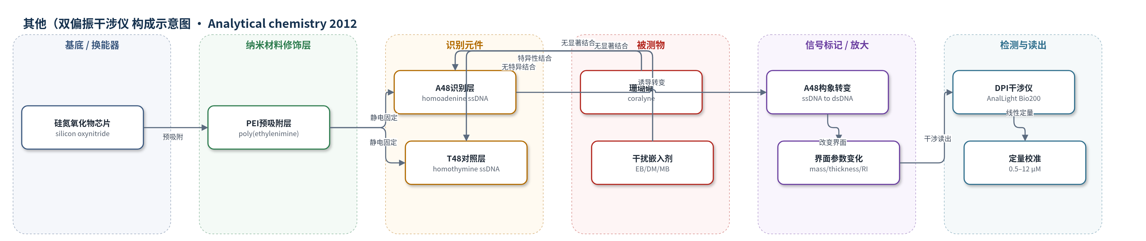
点击图片查看大图 · 依据论文自动绘制
传感器类型
其他(双偏振干涉仪(DPI)生物传感器)
检测对象
珊瑚碱(coralyne);样品基质:PBS缓冲液(10 mM PBS, pH 7.4, 150 mM NaCl)
检测原理
硅氮氧化物DPI芯片表面先预吸附PEI形成正电荷层,再静电固定A48。当溶液中的coralyne与A48特异性结合时,平面碱性小分子嵌入A48并诱导腺嘌呤-腺嘌呤碱基配对,使A48由单链松散结构转变为更厚、更刚性的双链样自组装结构。该结合与构象转变引起传感界面质量增加、厚度增加和折射率降低。DPI利用TM和TE偏振光在多层波导中形成干涉,实时解析表面层的质量、厚度和折射率。由于coralyne与A48的结合可逆,信号随浓度升高而增大,在0.5–12 μM范围内质量、厚度和RI变化与浓度呈线性关系,从而实现无标记定量检测。
检测灵敏度
LOD (S/N=3): 0.22 μM for mass calibration, 0.14 μM for thickness calibration, 0.32 μM for RI calibration;线性范围: 0.5–12 μM;灵敏度斜率: M = 0.019c − 0.017 (ng mm−2), T = 0.09c − 0.11 (nm), RI = −0.0016c + 0.0013 (c单位μM);相关系数: r = 0.993 (mass), r = 0.993 (thickness), r = 0.990 (RI)
效应效果
该DPI传感器对coralyne选择性高:T48/PEI和裸PEI对照界面在0.5–12 μM下质量、厚度和RI变化明显小于A48/PEI;溴化乙锭(EB)、柔红霉素(DM)和亚甲基蓝(MB)均未引起明显变化,说明响应来自A48对coralyne的特异性识别。0.5–12 μM内质量、厚度和RI响应线性,检测限分别为0.22、0.14和0.32 μM,作者认为与金/银纳米颗粒比色法相当。结合曲线给出ka=4.95×10^3 M−1 s−1、kd=0.031 s−1、KA=1.6×10^5 M−1,可实时获得动力学信息。论文未报告RSD、稳定性或实际样品回收率,但提出可拓展用于多种靶分子与功能DNA相互作用检测。
传感器的构成
- 基底/换能器:硅氮氧化物AnalChip(silicon oxynitride AnalChip),DPI光学波导芯片,提供传感表面并产生干涉信号
- 预吸附修饰层:聚乙烯亚胺(PEI, MW=750000),在芯片表面形成正电荷层,用于静电吸附ssDNA
- 识别元件:48-mer均聚腺嘌呤单链DNA(A48, homoadenine ssDNA),特异性结合coralyne并诱导构象转变
- 对照识别元件:48-mer均聚胸腺嘧啶单链DNA(T48, homothymine ssDNA),用于排除非特异结合
- 信号读出:双偏振干涉仪(DPI, AnalLight Bio200),通过TM/TE偏振干涉实时解析质量、厚度和折射率变化
中文摘要
我们将腺嘌呤富集单链DNA(ssDNA)的特异性识别引入双偏振干涉仪(DPI)测量,实现小分子珊瑚碱(coralyne)的直接、选择性和灵敏检测,并利用该实时无标记技术研究珊瑚碱与腺嘌呤富集ssDNA的相互作用。紫外-可见光谱、圆二色光谱和DNA熔解实验证实,48-mer均聚腺嘌呤ssDNA寡核苷酸(A48)对珊瑚碱具有高度特异性识别,而48-mer均聚胸腺嘧啶ssDNA(T48)无此识别。ssDNA通过预吸附聚乙烯亚胺(PEI)层固定在硅氮氧化物芯片上。DPI解析的质量、厚度和折射率(RI)变化表明,ssDNA主要以平躺单层形式位于PEI表面并部分插入PEI层。加入不同浓度珊瑚碱后,A48/PEI层的质量、厚度和RI变化揭示A48可能由单链结构转变为双链结构。由质量结合曲线求得ka、kd和KA分别为4.95×10^3 M^-1 s^-1、0.031 s^-1和1.6×10^5 M^-1。在0.5–12 μM范围内,珊瑚碱浓度与质量、厚度和RI响应线性相关;检测限分别为0.22 μM、0.14 μM和0.32 μM。T48/PEI、PEI界面及溴化乙锭、柔红霉素、亚甲基蓝对照证实了高选择性。
英文摘要
We incorporated the specific recognition of adenine-rich singled-stranded DNA (ssDNA) into dual polarization interferometry (DPI) measurements for direct, selective, and sensitive detection of the small molecule coralyne, and we simultaneously employed the real-time and label-free technique for detailed investigation of the interaction between coralyne and adenine-rich ssDNA. Data from UV-visible spectroscopy, circular dichroism (CD) spectroscopy, and DNA melting firmly confirmed that 48-mer homoadenine ssDNA oligonucleotide (A(48)) had highly specific recognition for coralyne, whereas 48-mer homothymine ssDNA oligonucleotide (T(48)) as the control had no such recognition. The immobilization of ssDNA (A(48) or T(48)) on a silicon oxynitride chip could be achieved through a preadsorbed poly(ethylenimine) (PEI) layer. Mass, thickness, and refractive index (RI) changes resolved by DPI during the whole process of ssDNA immobilization suggested that most ssDNA molecules were likely to lie on the PEI surface mainly in the form of a flat monolayer and insert themselves partly into the PEI layer. Qualitative and quantitative analysis of mass, thickness, and RI changes in A(48)/PEI layer upon addition of different concentrations of coralyne revealed that A(48) most likely underwent a conformational change from single-stranded to double-stranded structure. By evaluation of the binding curves from changes in mass, the association rate constant (k(a)), dissociation rate constant (k(d)), and association constant (K(A)) between coralyne and A(48) were determined to be 4.95 × 10(3) M(-1) s(-1), 0.031 s(-1), and 1.6 × 10(5) M(-1), respectively. Good linear correlations between coralyne concentrations ranging from 0.5 to 12 μM and three parameters (mass, thickness, and RI) resolved by the response to coralyne binding were obtained. The detection limits were 0.22 μM for mass calibration, 0.14 μM for thickness calibration, and 0.32 μM for RI calibration. The high selectivity of the biosensor to coralyne at the A(48)/PEI interface was successfully confirmed by using the other two interfaces (T(48)/PEI and PEI) and three typical intercalators (ethidium bromide, daunomycin, and methylene blue). It is expected that the biosensing platform may be extended to simultaneously detect and characterize the interactions of a variety of target molecules with functional DNA molecules with high sensitivity.