组成图示
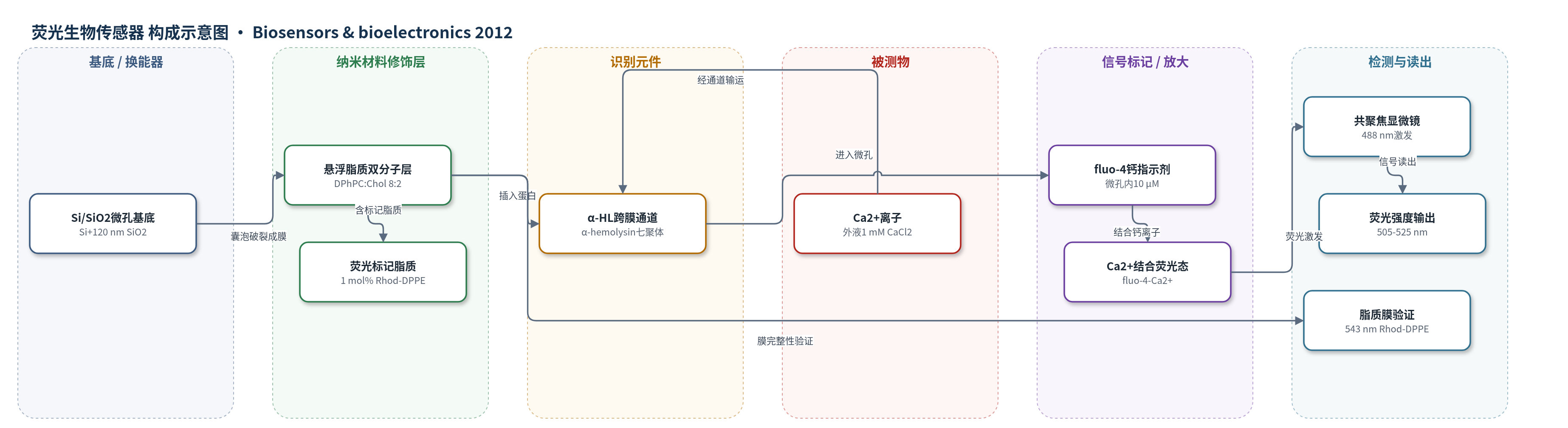
点击图片查看大图 · 依据论文自动绘制
传感器类型
荧光生物传感器
检测对象
钙离子(Ca2+)跨膜输运/α-溶血素(α-hemolysin, α-HL)通道活性;样品基质:人工脂质双分子层微孔阵列中的160 mM葡萄糖、20 mM NaCl、1 mM EDTA缓冲液及含1 mM CaCl2外液
检测原理
该传感器以人工脂质双分子层密封的硅微孔为传感单元。α-溶血素(α-HL)从外液扩散至悬空脂质膜,插入膜中并七聚形成跨膜孔道;外液加入CaCl2至1 mM,使微孔内外形成Ca2+浓度梯度,Ca2+沿梯度经α-HL通道进入初始无钙的微孔。微孔内预封闭的fluo-4与进入的Ca2+结合,荧光强度增强;微孔体积仅1–100 fL,使极小的离子通量即可引起可测的局部浓度变化,从而实现信号放大。α-HL浓度越高,膜中通道数越多,荧光上升越快并更早达到饱和。共聚焦显微镜以488 nm激发fluo-4,收集505–525 nm发射,将Ca2+输运速率转换为荧光强度时间曲线。
检测灵敏度
LOD: several tens of ions/s/μm2
效应效果
该装置在人工脂质双分子层微孔阵列中实现Ca2+输运监测,微孔体积1–100 fL,检测限为数十ions/s/μm2,比标准电生理测量低4–5个数量级。脂质膜稳定,fluo-4泄漏可忽略,荧光基线稳定;Ca2+荧光上升速率约为fluo-4泄漏下降速率的20倍。不同微孔响应起始时间存在约2 min差异,2 μm与4 μm微孔的fluo-4泄漏平均衰减率分别为0.63±0.04和0.72±0.02 ×10^-3/s,对应12和54 molecules/s。α-HL浓度100 nM–1 μM时,通道形成概率和输运速率随浓度升高而增加,1 μM时约80 s达最大荧光。作者认为该阵列可扩展至其他离子指示剂,实现多离子并行检测,是膜蛋白功能分析和高通量纳米生物器件的基础组件。
传感器的构成
- 基底/微孔结构:Si基底+120 nm SiO2热氧化层,光刻与KOH刻蚀形成1/2/4/8 μm直径、1 μm深微孔及overhang,支撑脂质膜并封闭探针
- 脂质双分子层:DPhPC与胆固醇(8:2)电形成GUV破裂形成悬浮人工脂质双分子层,作为跨膜屏障并承载通道蛋白
- 标记脂质:1 mol% Rhod-DPPE掺入脂质膜,543 nm激发下验证脂质膜完整性
- 识别/功能元件:α-hemolysin(α-HL)插入脂质膜并七聚形成跨膜孔道,允许Ca2+和fluo-4输运
- 信号标记物:fluo-4(10 μM)封闭在微孔内,与Ca2+结合后荧光增强
- 缓冲/离子环境:160 mM葡萄糖、20 mM NaCl、1 mM EDTA填充微孔,外液加CaCl2至1 mM,维持渗透压并提供Ca2+浓度梯度
- 光学读出:共聚焦激光扫描显微镜488 nm激发、505–525 nm带通滤片检测fluo-4荧光
中文摘要
为实现离子通道活性的功能分析,在硅基底上通过破裂巨单层囊泡形成悬置于微孔上方的人工脂质双分子层。钙离子指示剂fluo-4被脂质双分子层密封封闭在微孔内;微孔口形成的悬挑结构可防止脂质膜落入孔内,从而实现荧光探针的稳定封闭。将α-溶血素插入脂质膜形成通道后,通过监测微孔内fluo-4的荧光强度变化,分析Ca2+经通道的跨膜输运。微孔体积很小(1–100 fL),因此可实现高灵敏监测,检测限为数十个离子/s/μm2,远小于标准电生理测量中的离子电流。更小的微孔有望模拟细胞内局部离子浓度变化,但单通道功能分析仍需进一步提高信噪比。结果表明,由脂质双分子层密封并封闭荧光探针的微孔阵列可构成基于功能性膜蛋白的高灵敏生物传感器阵列的基本单元,有望实现高通量、平行化检测装置。
英文摘要
For the functional analysis of ion channel activity, an artificial lipid bilayer suspended over microwells was formed that ruptured giant unilamellar vesicles on a Si substrate. Ca(2+) ion indicators (fluo-4) were confined in the microwells by sealing the microwells with a lipid bilayer. An overhang formed at the microwells prevented the lipid membrane from falling into them and allowed the stable confinement of the fluorescent probes. The transport of Ca(2+) ions through the channels formed by α-hemolysin inserted in a lipid membrane was analyzed by employing the fluorescence intensity change of fluo-4 in the microwells. The microwell volume was very small (1-100 fl), so a highly sensitive monitor could be realized. The detection limit is several tens of ions/s/μm(2), and this is much smaller than the ion current in a standard electrophysiological measurement. Smaller microwells will make it possible to mimic a local ion concentration change in the cells, although the signal to noise ratio must be further improved for the functional analysis of a single channel. We demonstrated that a microwell array with confined fluorescent probes sealed by a lipid bilayer could constitute a basic component of a highly sensitive biosensor array that works with functional membrane proteins. This array will allow us to realize high throughput and parallel testing devices.