组成图示
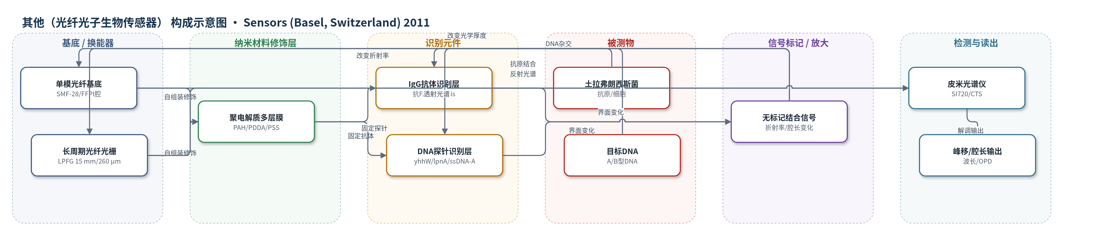
点击图片查看大图 · 依据论文自动绘制
传感器类型
其他(光纤光子生物传感器)
检测对象
土拉弗朗西斯菌(Francisella tularensis, F. tularensis)、F. tularensis subsp. tularensis(A型,TI0902)、F. tularensis subsp. holarctica(B型,LVS)、F. tularensis目标DNA(yhhW/lpnA/ssDNA-B);样品基质:辐照细胞悬液、细菌裂解上清、均相缓冲液
检测原理
该传感器采用无标记光子换能机制。LPFG免疫传感中,LbL/ESA聚电解质膜将IgG固定于光纤包层表面;F. tularensis抗原与IgG结合后,界面薄膜质量与折射率增加,改变LPFG芯模与包层模耦合条件,使透射光谱谐振峰向短波方向移动,峰波长降低量随抗原量增加。FFPI核酸传感中,DNA探针固定于光纤端面,目标DNA杂交或抗原结合使端面薄膜光学厚度增加,多腔干涉反射光谱发生频移;通过矩阵光学模型解调光纤腔/空气腔长度,可分辨0.1 nm变化。多腔结构提供内置温度补偿。整个体系无需PCR、荧光或酶标记,信号随目标DNA或抗原浓度增加而增大。
检测灵敏度
LOD: 约1 ng(目标DNA);线性范围: 1.7–163.8 ng(目标DNA);LOD: 至少10^5 CFU(F. tularensis细胞)
效应效果
LPFG传感器在生理盐水中无峰移,可检测至少10^5 CFU的F. tularensis细胞。FFPI信号处理可分辨0.1 nm腔厚变化,并在重复实验中显示稳定一致的探针固定。特异性方面,100%同源目标DNA产生4.2 nm正腔厚增加;含2 bp、5 bp、10 bp错配序列及随机阴性对照在128–152 µM下未产生正变化。亚种区分中,schu4-330探针仅使A型TI0902腔厚增加,lpnA探针使A型和B型LVS均增加,且lpnA对TI0902响应大于LVS。方法无需PCR,可检测纳克级DNA,作者认为其快速、免培养、可复用、低成本,适合现场诊断。
传感器的构成
- 基底/换能器:单模光纤(Corning SMF-28)、长周期光纤光栅(LPFG,15 mm,周期260 µm)和多腔光纤法布里-珀罗干涉仪(FFPI,含空气腔与光纤腔),作为光波导与谐振/干涉换能器。
- 纳米材料修饰层:聚电解质多层膜(LbL/ESA),由聚烯丙基胺盐酸盐(PAH)、聚二烯丙基二甲基氯化铵(PDDA)和聚苯乙烯磺酸钠(PSS)交替沉积,形成纳米结构薄膜,用于固定识别元件并调节界面折射率/光学厚度。
- 识别元件(免疫):抗F. tularensis血清IgG抗体,经Protein A亲和层析纯化,自组装固定于LPFG表面,用于捕获土拉弗朗西斯菌抗原。
- 识别元件(核酸):单链DNA探针,包括yhhW/schu4-330(101 bp,A型特异)、lpnA(117 bp,A/B型共有)及26-mer ssDNA-A探针,通过LbL/ESA固定于FFPI端面,用于目标DNA杂交。
- 可选封闭剂:PBS-1% BSA-0.1% NaN3,用于抑制非特异结合;原文指出若需要可作为最终制备步骤,本实验未额外使用。
- 信号标记物:无标记(label-free)体系,不使用荧光、酶或化学发光标记;识别事件直接改变薄膜折射率或光学厚度。
- 读出系统:高速皮米光谱仪/组件测试系统(Micron Optics SI720/CTS,1520–1570 nm)、光纤环形激光器与光纤环行器,监测透射/反射光谱峰移或腔长变化。
中文摘要
本研究探讨光子生物传感器检测A类病原体土拉弗朗西斯菌(Francisella tularensis)的应用。成功演示干涉型和长周期光纤光栅(LPFG)两种结构,均可检测免疫结合或DNA杂交引起的光学变化。通过逐层静电自组装(LbL/ESA)将DNA探针或IgG直接固定于光纤表面。采用15 mm、周期260 µm的透射式LPFG并涂覆抗F. tularensis血清IgG;IgG形成纳米薄膜,用光谱椭偏仪测折射率变化,通过抗原结合导致峰波长降低检测细菌。进一步用单模多腔光纤法布里-珀罗干涉仪(FFPI)区分A型TI0902和B型LVS。传感器在光纤端面沉积七层聚合物双分子层后固定yhhW(101 bp,A型特异)或lpnA(117 bp,A/B共有)探针。yhhW仅与A型反应,lpnA与A、B型均反应。方法可检测纳克级目标DNA,无需PCR,对100%同源DNA有反应,对2 bp错配无反应,特异性高,有望用于快速、免培养、现场诊断。
英文摘要
The application of photonic biosensor assays to diagnose the category-A select agent Francisella tularensis was investigated. Both interferometric and long period fiber grating sensing structures were successfully demonstrated; both these sensors are capable of detecting the optical changes induced by either immunological binding or DNA hybridization. Detection was made possible by the attachment of DNA probes or immunoglobulins (IgG) directly to the fiber surface via layer-by-layer electrostatic self-assembly. An optical fiber biosensor was tested using a standard transmission mode long period fiber grating of length 15 mm and period 260 μm, and coated with the IgG fraction of antiserum to F. tularensis. The IgG was deposited onto the optical fiber surface in a nanostructured film, and the resulting refractive index change was measured using spectroscopic ellipsometry. The presence of F. tularensis was detected from the decrease of peak wavelength caused by binding of specific antigen. Detection and differentiation of F. tularensis subspecies tularensis (type A strain TI0902) and subspecies holarctica (type B strain LVS) was further accomplished using a single-mode multi-cavity fiber Fabry-Perot interferometric sensor. These sensors were prepared by depositing seven polymer bilayers onto the fiber tip followed by attaching one of two DNA probes: (a) a 101-bp probe from the yhhW gene unique to type-A strains, or (b) a 117-bp probe of the lpnA gene, common to both type-A and type-B strains. The yhhW probe was reactive with the type-A, but not the type-B strain. Probe lpnA was reactive with both type-A and type-B strains. Nanogram quantities of the target DNA could be detected, highlighting the sensitivity of this method for DNA detection without the use of PCR. The DNA probe reacted with 100% homologous target DNA, but did not react with sequences containing 2-bp mismatches, indicating the high specificity of the assay. These assays will fill an important void that exists for rapid, culture-free, and field-compatible diagnosis of F. tularensis.