组成图示
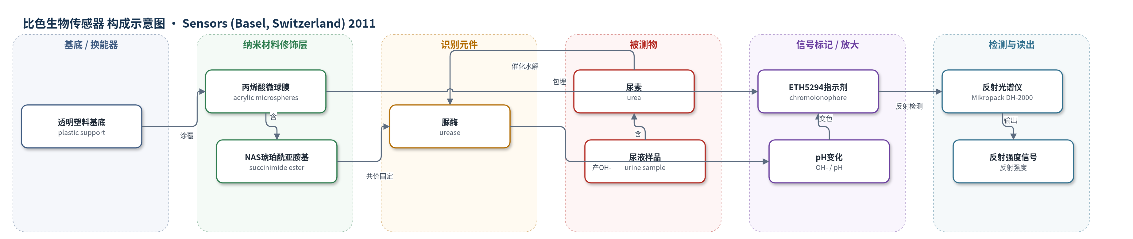
点击图片查看大图 · 依据论文自动绘制
传感器类型
比色生物传感器
检测对象
尿素(urea);样品基质:尿液(urine),亦使用磷酸盐缓冲液(phosphate buffer)
检测原理
传感器以脲酶为识别元件,以pH敏感色离子指示剂ETH 5294为信号元件。当尿液或缓冲液中的尿素扩散到固定化脲酶表面时,脲酶催化尿素水解生成氨和碳酸,氨在水中进一步生成NH4+和OH−,使微球界面pH升高。ETH 5294的质子化状态随pH改变而发生颜色变化,改变微球膜对入射光的反射特性。反射分光光度计读取反射强度变化,尿素浓度越高,pH升高越明显,指示剂颜色变化越大,反射强度变化越大。该过程依靠酶催化实现化学放大,微球大比表面积和反射率换能有助于获得宽线性范围。
检测灵敏度
LOD: 9.97 μM;线性范围: 0.01–1,000 mM;灵敏度: 146.99 I/mM (pH 6.5);R^2 = 0.97
效应效果
在pH 6.5磷酸盐缓冲液中,传感器对10.0 mM尿素的反射响应重现性良好,RSD为1.43%(n=5)。对Na+、K+、NH4+和Mg2+(0.001–100 mM)无明显干扰,表明对尿素具有较好选择性。4 °C保存45 d后,1.0和10.0 mM尿素的反射强度仍保持初始强度的87.01%和82.37%,稳定性优于部分溶胶-凝胶脲酶传感器。尿液加标回收率为94.91%–118.73%;与DMAB化学法比较,相关系数R2=0.997、斜率1.18。作者认为其宽线性范围、低检出限和良好稳定性适合尿液尿素检测。
传感器的构成
- 基底:透明塑料片(plastic support),承载微球膜并提供反射光学基底
- 微球基质:丙烯酸微球(acrylic microspheres),由n-BA、HDDA、DMPP、NAS和SDS光聚合制备,提供大比表面积并包埋指示剂
- 功能化修饰层:N-丙烯酰琥珀酰亚胺(NAS)琥珀酰亚胺酯基团,接枝于微球表面,作为脲酶共价连接位点
- 识别元件:脲酶(urease,E.C. 3.5.1.5),共价固定于微球表面,催化尿素水解
- 信号标记物:尼罗蓝色离子指示剂ETH 5294,分散/包埋于丙烯酸微球中,随pH变化发生颜色变化
- 读出装置:反射分光光度计(reflectance spectrophotometer,Mikropack DH-2000-Bal),检测颜色变化引起的反射强度变化
中文摘要
本研究通过光聚合合成新型丙烯酸微球,并在微球制备过程中引入琥珀酰亚胺功能基团。利用该材料成功构建了基于反射率换能、对尿素具有宽线性响应范围的光学生物传感器。传感器采用琥珀酰亚胺修饰的丙烯酸微球,其中包埋尼罗蓝色离子指示剂ETH 5294用于光学检测,并通过微球表面的琥珀酰亚胺基团共价固定脲酶。实验表明酶和指示剂均无渗漏。脲酶水解尿素使体系pH升高,导致固定化色离子指示剂发生颜色变化;以反射分光光度法监测该颜色变化时,传感器对尿素的线性响应范围为0.01–1000 mM(R2=0.97),检出限为9.97 μM。传感器重现性良好(RSD=1.43%,n=5),且Na+、K+、NH4+和Mg2+等主要阳离子无明显干扰。采用反射率作为换能方式可获得比许多其他光学换能尿素生物传感器更大的线性响应范围。
英文摘要
New acrylic microspheres were synthesised by photopolymerisation where the succinimide functional group was incorporated during the microsphere preparation. An optical biosensor for urea based on reflectance transduction with a large linear response range to urea was successfully developed using this material. The biosensor utilized succinimide-modified acrylic microspheres immobilized with a Nile blue chromoionophore (ETH 5294) for optical detection and urease enzyme was immobilized on the surface of the microspheres via the succinimide groups. No leaching of the enzyme or chromoionophore was observed. Hydrolysis of the urea by urease changes the pH and leads to a color change of the immobilized chromoionophore. When the color change was monitored by reflectance spectrophotometry, the linear response range of the biosensor to urea was from 0.01 to 1,000 mM (R2 = 0.97) with a limit of detection of 9.97 μM. The biosensor response showed good reproducibility (relative standard deviation = 1.43%, n = 5) with no interference by major cations such as Na+, K+, NH4+ and Mg2+. The use of reflectance as a transduction method led to a large linear response range that is better than that of many urea biosensors based on other optical transduction methods.