组成图示
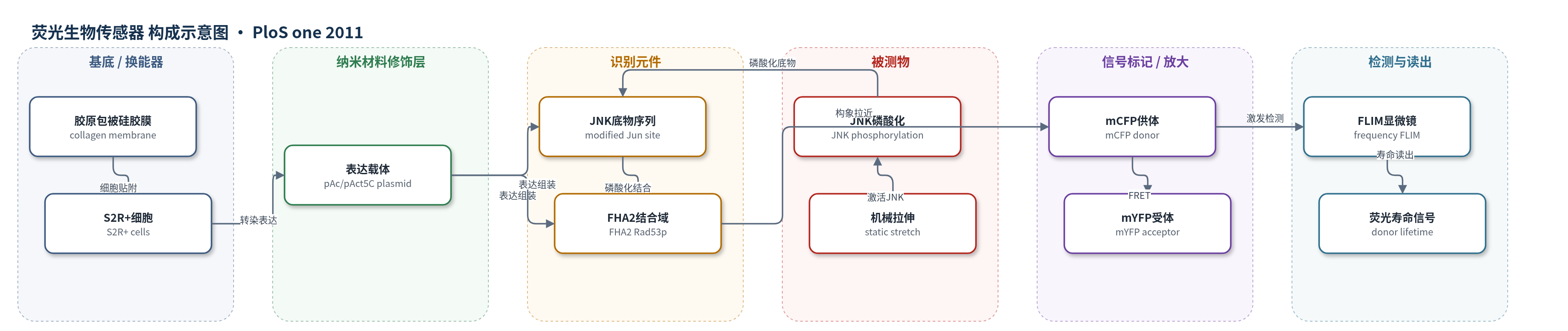
点击图片查看大图 · 依据论文自动绘制
传感器类型
荧光生物传感器
检测对象
JNK通路活性(JNK signaling pathway activity)、JNK介导的c-Jun磷酸化(JNK-mediated c-Jun phosphorylation);样品基质:活体果蝇S2R+细胞(Drosophila S2R+ cells)
检测原理
该传感器为基因编码FRET探针。机械拉伸通过细胞表面整合素和黏着斑激活JNK通路,JNK磷酸化dJun-FRET中的Jun底物序列。磷酸化序列被FHA2磷酸苏氨酸结合域识别并结合,引发分子内夹钳构象变化,使mCFP供体与mYFP受体距离缩短,FRET效率升高。FRET增强导致mCFP荧光寿命缩短。频域FLIM通过调制激发光测量mCFP寿命,寿命降低反映JNK活性升高。该过程无需外源标记或酶放大,信号随JNK磷酸化水平变化;2.5%静态拉伸可在20分钟内引起寿命下降,约2小时达到平台。
检测灵敏度
原文未报告LOD、线性范围、灵敏度斜率或相关系数。
效应效果
dJun-FRET对LPS和L-JNKI1响应明显,EGF无影响,显示选择性。FLIM不受激发强度和浓度影响,避免串扰。静态拉伸使胶原膜细胞寿命从2.43±0.15 ns降至1 h 2.18±0.15 ns、2 h 2.01±0.15 ns,并稳定数小时。基底改变基线:玻璃1.98±0.14 ns、胶原玻璃2.24±0.14 ns、Con-A玻璃2.19±0.15 ns。Mys RNAi使拉伸无法激活(2.21±0.11至2.23±0.11 ns),talin RNAi仍可激活(2.10±0.13至1.98±0.12 ns)。受体光漂白后寿命由1.97±0.15 ns恢复至2.34±0.12 ns,验证FRET。作者认为适合活细胞实时、高通量JNK动态分析。
传感器的构成
- 细胞培养基底:胶原包被硅胶膜(collagen-coated silicone membrane)、玻璃或塑料,提供细胞附着与机械拉伸形变
- 表达载体:pAc/pAct5C质粒,驱动dJun-FRET传感器在Drosophila S2R+细胞中表达
- 识别元件:JNK底物序列(modified Jun phosphorylation site)与FHA2磷酸苏氨酸结合域(FHA2, Rad53p),识别JNK介导的磷酸化
- 信号标记物:mCFP供体与mYFP受体荧光蛋白,通过FRET报告传感器构象变化
- 换能读出:频域荧光寿命成像系统(FLIM, LIFA),检测mCFP荧光寿命变化
中文摘要
机械力已知可调节Jun N端激酶(JNK)信号级联,但此前对机械应力激活JNK的分析主要依赖体外检测方法,活细胞中JNK如何响应机械应力及其功能仍不清楚。本研究利用荧光寿命成像显微镜(FLIM)和分子内磷酸化依赖的dJun-FRET生物传感器,实时评估果蝇S2R+细胞中JNK通路活性。定量FRET-FLIM分析与共聚焦显微镜显示,机械拉伸可诱导dJun-FRET传感器持续激活,并伴随稳定的细胞形态变化。培养在不同基底上的细胞表现出不同水平的JNK活性,且与细胞形态、整合素表达和黏着斑组织差异相关。结果表明,细胞骨架和基质附着改变可调节JNK信号,JNK活性也可能反馈调节细胞骨架与细胞黏附。静息状态下,黏着斑中的整合素和talin是抑制JNK活性的关键因素;多向静态拉伸则引起整合素依赖、可能不依赖talin的Jun传感器激活。数据提示JNK活性需与其他信号元件协调,以调控拉伸相关的细胞骨架和细胞形状重塑。
英文摘要
Mechanical force is known to modulate the activity of the Jun N-terminal kinase (JNK) signaling cascade. However, the effect of mechanical stresses on JNK signaling activation has previously only been analyzed by in vitro detection methods. It still remains unknown how living cells activate the JNK signaling cascade in response to mechanical stress and what its functions are in stretched cells.We assessed in real-time the activity of the JNK pathway in Drosophila cells by Fluorescence Lifetime Imaging Microscopy (FLIM), using an intramolecular phosphorylation-dependent dJun-FRET (Fluorescence Resonance Energy Transfer) biosensor. We found that quantitative FRET-FLIM analysis and confocal microscopy revealed sustained dJun-FRET biosensor activation and stable morphology changes in response to mechanical stretch for Drosophila S2R+ cells. Further, these cells plated on different substrates showed distinct levels of JNK activity that associate with differences in cell morphology, integrin expression and focal adhesion organization.These data imply that alterations in the cytoskeleton and matrix attachments may act as regulators of JNK signaling, and that JNK activity might feed back to modulate the cytoskeleton and cell adhesion. We found that this dynamic system is highly plastic; at rest, integrins at focal adhesions and talin are key factors suppressing JNK activity, while multidirectional static stretch leads to integrin-dependent, and probably talin-independent, Jun sensor activation. Further, our data suggest that JNK activity has to coordinate with other signaling elements for the regulation of the cytoskeleton and cell shape remodeling associated with stretch.