组成图示
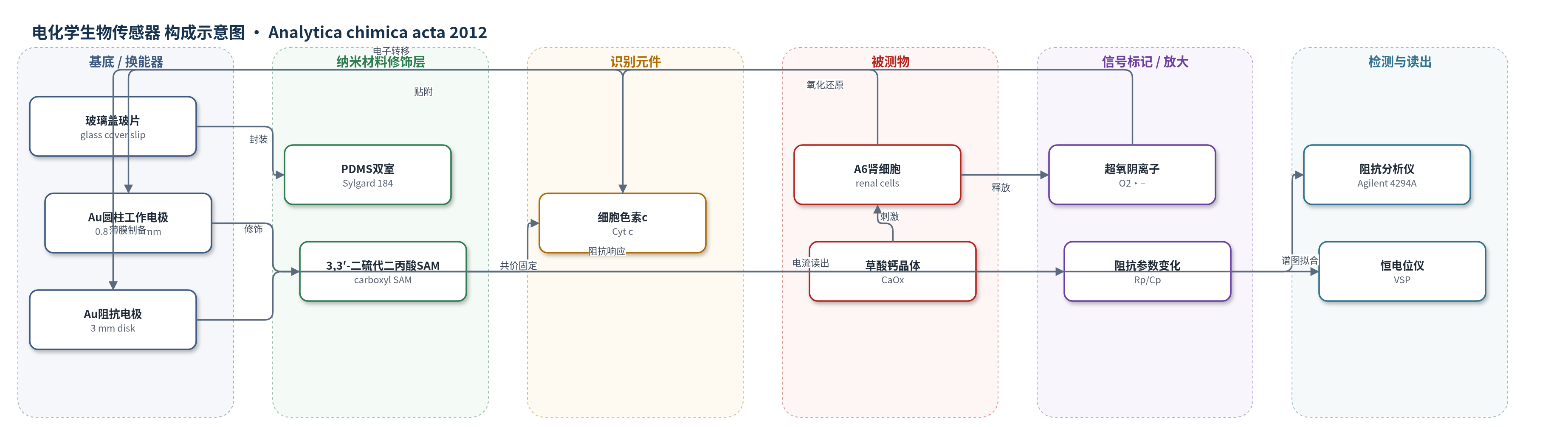
点击图片查看大图 · 依据论文自动绘制
传感器类型
电化学生物传感器
检测对象
超氧阴离子(superoxide anion, O2•−)、细胞贴附/跨上皮电阻(cell adherence/transepithelial resistance);样品基质:A6 肾上皮细胞培养体系(细胞外空间,草酸钙晶体刺激)
检测原理
系统由阻抗通道与安培通道组成。阻抗通道向 Au 电极施加零偏 20 mV RMS 交流信号(400 Hz–600 kHz),细胞膜电容与跨上皮电阻构成等效电路;草酸钙引起细胞肿胀、贴附减弱或脱离时,CPE/Rp/Cp 等参数实时变化。安培通道中,Cyt c 经 SAM 共价固定于 Au 工作电极,在 150 mV 下催化细胞释放的 O2•− 发生电子转移,产生与 O2•− 浓度成正比的电流;其线性范围可达 1 µM,灵敏度为 18.3±2.7×10^2 A m−2 M−1。两通道同步读出,实现贴附与氧化应激的实时关联分析。
检测灵敏度
线性范围: up to 1 µM superoxide;灵敏度: 18.3 ± 2.7 × 10^2 A m−2 M−1
效应效果
0.91 mg/mL 草酸钙刺激约 1 h,阻抗拟合 chi-squared 为 0.00032–0.00058;加草酸钙后 CPE cells-T 增 33%,CPE cells-P 降 4%,R cells 降 10%,电极/溶液参数变化最大 2%。Cyt c 传感器对 H2O2、抗坏血酸、尿酸干扰小,但对 NO/过氧亚硝酸盐选择性未验证。玻璃微球可产生类似阻抗变化但无显著超氧阴离子电流,提示草酸钙兼具机械与化学损伤。亚汇合细胞比汇合细胞更敏感,系统可用于肾结石机制及贴壁细胞多参数研究。
传感器的构成
- 阻抗通道基底:玻璃盖玻片(glass cover slip)与薄膜 Au 工作/对电极,承载 A6 细胞并进行非法拉第阻抗谱测量
- 阻抗通道微结构:PDMS(Sylgard 184)双室,每室 250 µL,隔离阻抗与安培测量并减少电串扰
- 安培通道换能电极:圆柱形 Au 工作电极(直径 0.8 mm、高 4 mm),作为超氧阴离子安培检测的换能器
- 自组装单分子层:3,3′-二硫代二丙酸(3,3′-dithiodipropionic acid)SAM,在 Au 表面提供羧基锚定位点
- 识别/催化元件:细胞色素 c(cytochrome c, Cyt c)经 EDC/NHS 共价固定于 SAM,介导超氧阴离子电子转移
- 参比/对电极:Ag/AgCl(3 M KCl)参比电极与 Pt 丝对电极,构成三电极安培体系
- 样品/刺激物:A6 肾上皮细胞与草酸钙(calcium oxalate, CaOx)晶体,产生细胞贴附变化与超氧阴离子释放
中文摘要
即使最简单的实验刺激也会引发复杂的细胞响应,因此需要同时监测更多细胞参数。本文建立了一种全电化学系统,可实时同步监测细胞贴附与细胞外超氧阴离子释放。细胞贴附(细胞与邻近细胞及基底之间的贴附)采用非法拉第阻抗谱监测;超氧阴离子释放采用基于细胞色素c的安培生物传感器监测。该系统首次用于实时观察暴露于成石盐草酸钙的肾细胞中上述两个参数的变化。结果表明,草酸钙晶体可降低细胞贴附,同时通过过量产生超氧阴离子诱导氧化应激。未形成完整紧密连接的亚汇合细胞比具有紧密连接的汇合细胞更脆弱,提示紧密连接具有重要保护作用。
英文摘要
The complexity of the cellular response, induced even by the simplest experimental stimulus, requires an increased number of cellular parameters to be simultaneously monitored. An all electrochemical system allowing the simultaneous and real-time monitoring of both cell adherence and superoxide release into the extracellular space was developed to address this challenge. Cell adherence (to neighboring cells and to substrate) was monitored using non-faradaic impedance spectroscopy while the superoxide release was monitored using a cytochrome c-based amperometric biosensor. The system was used to observe for the first time how these two cellular parameters are changing in real-time for renal cells exposed to calcium oxalate, a calculus-forming salt. It was discovered that calcium oxalate crystals decrease cell adherence and in the same time induce oxidative stress by an overproduction of superoxide. Subconfluent cells, without fully developed tight junctions, appear to be more vulnerable than confluent cells with tight junctions indicating the important protective role of these junctions.