组成图示
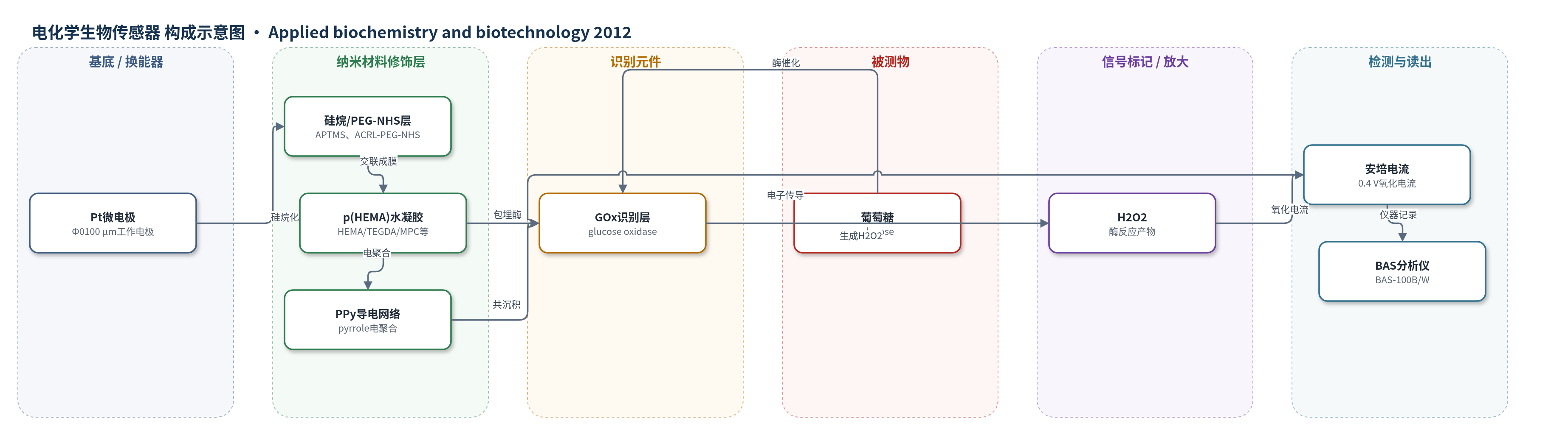
点击图片查看大图 · 依据论文自动绘制
传感器类型
电化学生物传感器
检测对象
葡萄糖(glucose, Glc);样品基质:100 mM PBKCl缓冲液(pH 7.0),非实际生物样品
检测原理
葡萄糖扩散进入p(HEMA)水凝胶/PPy互穿网络,被包埋的GOx催化氧化:β-D-葡萄糖与O2反应生成葡萄糖酸-δ-内酯和H2O2,GOx/FAD被还原后再被O2再生。H2O2扩散至Pt微电极表面,在0.4 V(vs. Ag/AgCl)发生氧化反应H2O2→O2+2H+ +2e−,产生与葡萄糖浓度相关的安培电流。PPy导电网络降低电极阻抗并筛选负电荷干扰物,水凝胶提供高水合环境以稳定GOx;过氧化的PPy增加膜致密性和扩散屏障,使响应受质量传输影响。通过Lineweaver–Burk分析稳态电流与葡萄糖浓度关系,获得KMapp和Imax,电流随葡萄糖浓度升高而增大。
检测灵敏度
LOD: 未报告;线性范围: 1.0 mC/cm2为1–8、1–10、1–15 mM,10.0 mC/cm2为0–7、0–7、1–10、1–15 mM;灵敏度: 1.0 mC/cm2为11.6、17.5、18.3、22.1、18.6、21.2 nA/mM,10.0 mC/cm2为6.4、3.6、8.5、10.6、12.8 pA/mM
效应效果
器件在4 °C PBKCl中储存至18 d,响应时间稳定为50 s,表明水凝胶与电极界面未明显脱层。1.0 mC/cm2膜灵敏度从11.6 nA/mM升至最高22.1 nA/mM,18 d为21.2 nA/mM,最大电流增至2.7 nA;10.0 mC/cm2膜灵敏度从6.4 pA/mM升至12.8 pA/mM,最大电流增至170 pA。10.0 mC/cm2的KMapp为2.7–6.1 mM,较1.0 mC/cm2的18.6–152.0 mM更稳定,说明更高PPy电荷密度有利于维持GOx亲和力,但增加扩散屏障并降低电流。未报告RSD、实际样品回收率及与ELISA/HPLC/qPCR对比。作者认为PPy电聚合可有效赋予水凝胶微电极生物活性,GOx活性随时间提高,但存在稳定性与电流响应的权衡。
传感器的构成
- 基底/换能器电极:Pt微电极(Φ0100 μm),经抛光、RCA清洗和电化学清洗,作为安培换能器
- 界面偶联层:3-aminopropyl-trimethoxysilane(APTMS)硅烷化引入氨基,ACRL-PEG-NHS(MW 400)引入丙烯酸酯基团,用于水凝胶共价固定
- 水凝胶层:poly(HEMA)-based hydrogel,含HEMA、TEGDA(3 mol%交联剂)、PEGMA、MPC、HMMA、p(NVP)、DMAEMA和DMPA光引发剂,UV交联约5 μm,提供高水合酶容纳环境
- 导电聚合物层:polypyrrole(PPy),由pyrrole电聚合形成,提供电子传导并筛选干扰物
- 识别元件:glucose oxidase(GOx,E.C. 1.1.3.4),包埋于PPy/水凝胶中,催化葡萄糖氧化
- 信号产物/检测介质:H2O2(酶反应产物)在0.4 V氧化产生电流;PBKCl(100 mM,pH 7.0)提供O2和离子导电环境
中文摘要
本研究通过电聚合过程赋予电极支撑水凝胶以酶生物特异性,形成由本征导电聚合物与高水合水凝胶组成的生物活性、刺激响应互穿网络。以聚(羟乙基甲基丙烯酸酯)基水凝胶包覆的Pt微电极(Φ0100 μm)为工作电极,在含吡咯(0.4 M)和葡萄糖氧化酶(GOx,1.0 mg/mL)的水溶液中,于750 mV(vs. Ag/AgCl,3 M KCl)恒电位电聚合吡咯,电荷密度分别为1.0和10.0 mC/cm2,制备葡萄糖响应生物换能器。随后在PBKCl(pH 7.0)中循环伏安过氧化聚吡咯(0–1.2 V,40圈)。器件在4 °C PBKCl中储存至18 d,并在0.4 V(vs. Ag/AgCl)进行安培剂量响应,经Lineweaver–Burk分析获得酶动力学参数。表观Michaelis常数KMapp在1.0 mC/cm2下由18.6增至152.0 mM,在10.0 mC/cm2下为2.7–6.1 mM;灵敏度分别增至21.2 nA/mM(18 d)和12.8 pA/mM(10 d),最大电流增至2.7 nA和170 pA。结果表明,聚吡咯电聚合可有效赋予水凝胶微电极生物活性,GOx在导电水凝胶中被稳定且活性随时间提高。
英文摘要
Electrode-supported hydrogels were conferred with the biospecificity of enzymes during the process of electropolymerization to give rise to a class of bioactive, stimuli-responsive co-joined interpenetrating networks of inherently conductive polymers and highly hydrated hydrogels. Glucose responsive biotransducers were prepared by potentiostatic electropolymerization [750 mV vs. Ag/AgCl (3 M KCl)] of pyrrole at Poly(hydoxyethyl methacrylate)-based hydrogel-coated Pt micro-electrodes (Φ = 100 μm) from aqueous solutions of pyrrole and glucose oxidase (GOx; 0.4 M pyrrole, 1.0 mg/ml GOx) to 1.0 and 10.0 mC/cm². Polypyrrole was them over-oxidized by cyclic voltammetry (0-1.2 V vs. Ag/AgCl, 40 cycles in PBKCl, pH = 7.0). Biotransducers were stored at 4 °C in PBKCl for up to 18 days. Amperometric dose-response at 0.4 V vs. Ag/AgCl followed by Lineweaver-Burk analysis produced enzyme kinetic parameters as a function of electropolymerization charge density and storage time. Apparent Michaelis constant (K (Mapp)) increased from 18.6-152.0 mM (1.0 mC/cm²) and from 2.7-6.1 mM (10.0 mC/cm²). Biotransducer sensitivity increased to 21.2 nA/mM after 18 days and to 12.8 pA/mM after 10 days for the 1.0 and 10.0 mC/cm² membranes, respectively. Maximum current, I (max), also increased over time to 2.7 nA (1.0 mC/cm²) and to 170 pA (10.0 mC/cm²). Electropolymerization of polypyrrole is shown to be an effective means for imparting bioactivity to a hydrogel-coated microelectrode. GOx was shown to be stabilized and to increase activity over time within the electroconductive hydrogel.