组成图示
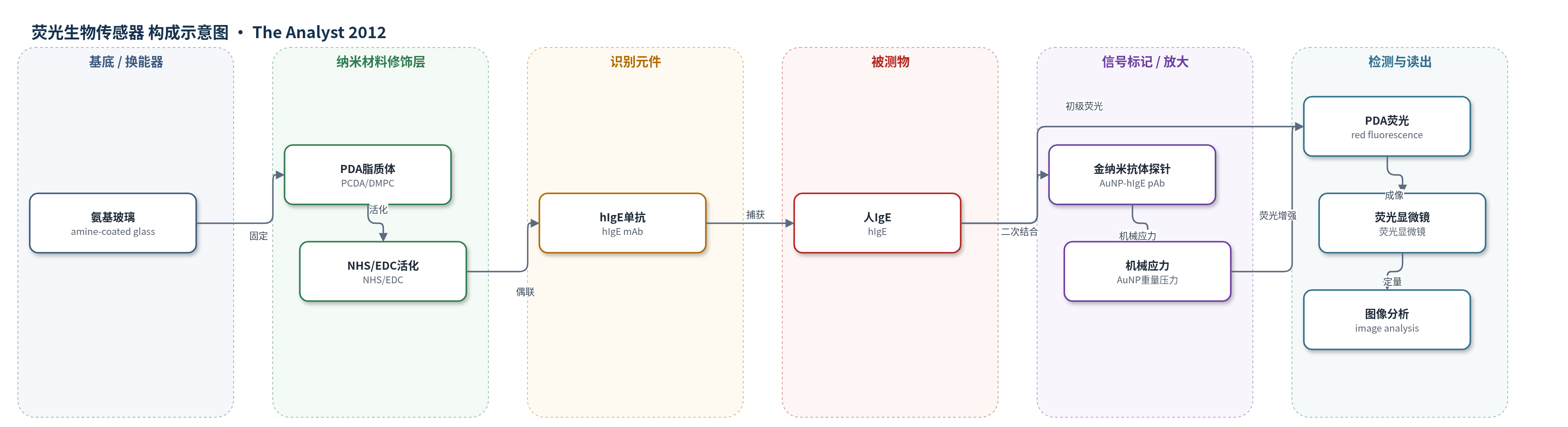
点击图片查看大图 · 依据论文自动绘制
传感器类型
荧光生物传感器
检测对象
人免疫球蛋白 E(human immunoglobulin E, hIgE);样品基质:PBS 缓冲液(pH 7.4),临床背景为血清
检测原理
该传感器以 PDA 脂质体为信号换能器。PDA 脂质体由 PCDA 与 DMPC 组成,经 254 nm 紫外光聚合形成共轭 ene–yne 骨架,未受扰时呈蓝色且荧光弱。当 hIgE 与固定在脂质体上的 hIgE 单克隆抗体结合时,抗原–抗体相互作用引起脂质体侧链和界面应力变化,使共轭骨架发生扰动,PDA 由蓝变红并产生红色荧光,荧光强度随 hIgE 浓度增加。进一步加入金纳米颗粒偶联 hIgE 多克隆抗体探针后,形成夹心免疫复合物;52 nm 金纳米颗粒因质量较大,对 PDA 脂质体施加额外机械压力,显著增强共轭骨架扰动和荧光发射。荧光显微镜采集斑点图像,经图像分析得到荧光强度,实现 hIgE 定量检测。
检测灵敏度
LOD: 10 ng mL−1(初级响应);0.1 ng mL−1(增强响应);线性范围: 10–10 000 ng mL−1(初级响应);0.1–10 000 ng mL−1(增强响应);灵敏度斜率: 9.527(初级响应);11.867(增强响应);R^2 = 0.970(初级响应);R^2 = 0.993(增强响应)
效应效果
该传感器在 PBS 中表现出良好选择性。1.0 mg/mL 的 BSA、纤维蛋白原和 hIgG 单独加入时,荧光信号仅轻微增加,与 PBS 本底相当,非特异吸附可忽略;在含 1.0 mg/mL 非靶蛋白和 1.0 mg/mL hIgE 的混合溶液中,hIgE 仍引起显著荧光增强,说明可在高浓度干扰蛋白存在下选择性检测目标蛋白。增强响应将 hIgE 最低可检测浓度从 10 ng/mL 降至 0.1 ng/mL,灵敏度提高约 100 倍,线性范围扩展至 0.1–10 000 ng/mL。文中未报告长期稳定性、实际血清样品加标回收率或与 ELISA/HPLC/qPCR 的直接对比。作者认为该策略可用于过敏诊断及超低浓度蛋白无标记检测。
传感器的构成
- 基底:氨基修饰玻璃(amine-coated glass),提供氨基用于固定 PDA 脂质体。
- 换能脂质体:PCDA/DMPC(8:2)PDA 脂质体,经 254 nm UV 聚合形成共轭 PDA 骨架,受刺激发生颜色与荧光变化。
- 活化交联层:NHS/EDC 活化 PCDA 羧基,乙二胺(ethylenediamine)交联脂质体,提高芯片稳定性。
- 识别元件:hIgE 单克隆抗体(hIgE mAb),经 NHS/EDC 偶联于 PDA 脂质体,特异性结合 hIgE。
- 信号放大元件:52 nm 羧基化金纳米颗粒(AuNPs,HS-OEG6-COOH/HS-OEG3-OH 封端)偶联 hIgE 多克隆抗体(hIgE pAb),二次结合后以重量/压力机械刺激 PDA 增强荧光。
- 读出系统:荧光显微镜(fluorescent microscopy)与图像分析软件,定量 PDA 斑点荧光强度。
中文摘要
聚二炔(PDA)脂质体具有对外界刺激产生颜色变化和荧光发射的独特性质,已被广泛研究作为生物传感器信号换能器。本文报道了一种基于 PDA 脂质体的抗体生物传感器,用于检测人免疫球蛋白 E(hIgE)。目标 hIgE 与固定在 PDA 脂质体上的 hIgE 单克隆抗体结合后,荧光信号随目标蛋白浓度略有增加,初级响应可检测至 10 ng/mL 以下。在初次免疫反应后加入金纳米颗粒偶联多克隆抗体探针,荧光信号随目标蛋白浓度显著增强。增强后的 PDA 脂质体生物传感器可将 hIgE 检出限降至 0.1 ng/mL,灵敏度较初级响应提高约 100 倍。结果表明,金纳米颗粒偶联多克隆抗体探针可有效增强 PDA 脂质体生物传感器芯片的荧光信号,该策略可用于超低浓度蛋白检测。
英文摘要
Polydiacetylene (PDA) liposomes possess unique properties that allow liposomes to change color and emit fluorescence in response to stimuli such as temperature, antibody-antigen interaction, pH, mechanical stress, and organic solvent. They have been studied extensively as signal transducers in biosensor applications. Here, we describe an antibody-based biosensor using PDA liposomes for detection of human immunoglobulin E (hIgE). Target hIgE chemically bound to hIgE monoclonal antibodies immobilized on PDA liposomes and the fluorescent signals were slightly increased depending on the target protein concentration. As the primary response, the hIgE could be detected to below 10 ng mL(-1). However, fluorescent signals were dramatically increased depending on the target protein concentration when gold nanoparticle-conjugated polyclonal antibody probes were added on the PDA liposomes after the primary immune reaction. A PDA liposome biosensor could detect the hIgE as low as 0.1 ng mL(-1) and the sensitivity was increased up to one hundred times higher than the primary response. As a result, we confirmed that gold nanoparticle-conjugated polyclonal antibody probes efficiently enhanced the fluorescent signal of the PDA liposome biosensor chip. This strategy can be useful to detect proteins of ultra-low concentration.