组成图示
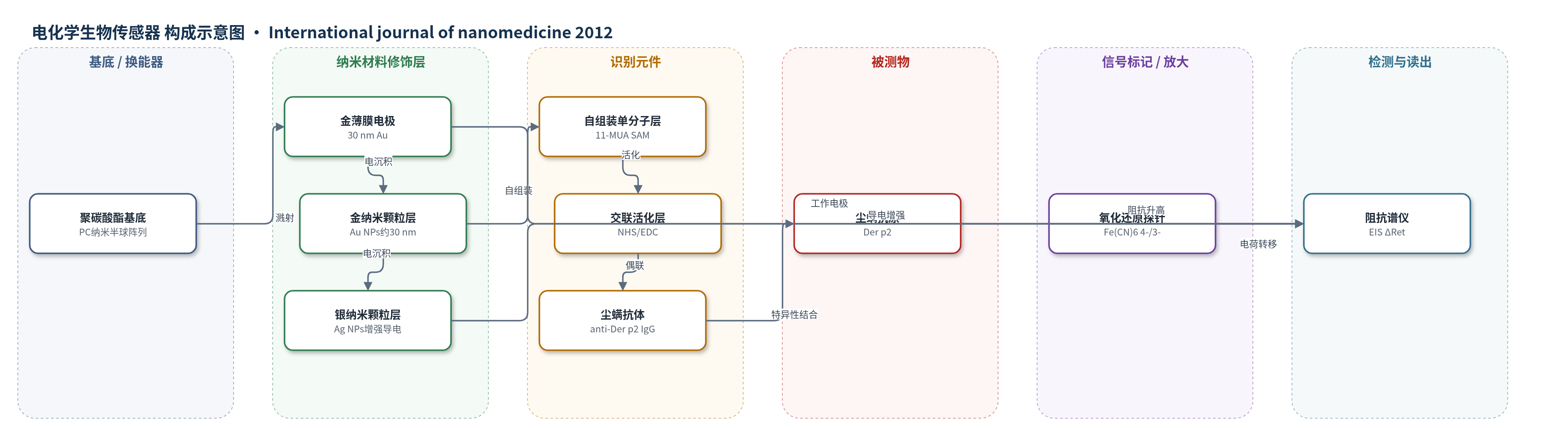
点击图片查看大图 · 依据论文自动绘制
传感器类型
电化学生物传感器
检测对象
尘螨抗原 Der p2(Dermatophagoides pteronyssinus group 2 allergen),样品基质为 Der p2 标准溶液(MOPS 缓冲液,pH 6.0)
检测原理
该传感器采用无标记电化学阻抗谱(EIS)检测。金纳米颗粒修饰的三维金电极增大有效面积,使更多11-MUA、NHS/EDC和抗Der p2 IgG负载于界面,提高识别位点密度。当Der p2与固定抗体特异性结合后,电极界面形成更厚的生物分子层,阻碍氧化还原探针Fe(CN)6 4-/3-在电极表面的电子转移,使电荷转移电阻Ret增大。EIS Nyquist图中半圆直径对应Ret,结合量越高,ΔRet越大。数据用Randles等效电路拟合,ΔRet与Der p2对数浓度呈二次关系,从而实现低浓度抗原定量检测。
检测灵敏度
LOD: 0.1 pg/mL(原文:around 0.1 pg/mL);动态范围: up to 10 ng/mL;R^2 = 0.98873;拟合式: y = -2,762x^2 -38,520x +17,538(x为log浓度,y为ΔRet)
效应效果
传感器以抗Der p2单克隆抗体作为识别元件,并用BSA封闭非特异位点,具有特异性识别能力。每个浓度重复三次,误差棒显示波动较小,但原文未报告RSD。电化学有效面积由0.043 cm2增至0.14 cm2,提高3.26倍,说明纳米结构增强了识别元件负载。检测限约0.1 pg/mL,动态范围至10 ng/mL,ΔRet与log浓度拟合R2=0.98873。作者认为该传感器低成本、一次性、高灵敏度,适合快速检测低丰度生物分子,未来可集成印刷电路板并配合便携式EIS设备。
传感器的构成
- 基底/换能器:纳米结构聚碳酸酯(PC)半球阵列基底,提供三维纳米形貌与机械支撑
- 电极层:射频磁控溅射30 nm金薄膜(Au thin film),作为工作电极
- 纳米修饰层:电化学沉积金纳米颗粒(Au NPs,平均约30±3 nm),增大有效电极面积并固定识别元件
- 导电增强层:电化学沉积银纳米颗粒(Ag NPs,含量约0.3%),提高电极导电性
- 识别固定层:11-巯基十二烷酸(11-MUA)自组装单分子层(SAM),提供羧基锚定
- 交联活化层:N-羟基琥珀酰亚胺(NHS)与碳二亚胺(EDC)活化羧基,用于共价偶联抗体
- 识别元件:尘螨单克隆抗体(IgG),特异性结合Der p2
- 封闭层:1%牛血清白蛋白(BSA),封闭非特异性结合位点
- 氧化还原探针:5 mM Fe(CN)6 4-/5 mM Fe(CN)6 3-(100 mM MOPS,pH 6.0),提供电荷转移阻抗信号
中文摘要
本研究整合纳米电铸、热压印和电化学沉积技术,开发一种一次性、低成本、高灵敏度的纳米结构生物传感器。以改性阳极氧化铝(AAO)阻挡层表面为模板沉积薄镍膜,刻蚀去除AAO模板后获得凹形纳米结构阵列的三维镍模;再利用该镍模通过热压印复制纳米结构聚碳酸酯(PC)基底。随后在PC基底上溅射薄金膜形成电极,并在三维金电极表面电化学沉积有序、均匀的金纳米颗粒(Au NPs)层;最后将银纳米颗粒(Ag NPs)沉积于金纳米颗粒上,以提高传感器导电性。采用电化学阻抗谱(EIS)检测目标物浓度,对尘螨抗原Der p2的检测灵敏度达到0.1 pg/mL。
英文摘要
This study integrates the techniques of nanoelectroforming, hot-embossing, and electrochemical deposition to develop a disposable, low-cost, and high sensitivity nanostructure biosensor. A modified anodic aluminum oxide barrier-layer surface was used as the template for thin nickel film deposition. After etching the anodic aluminum oxide template off, a three-dimensional mold of the concave nanostructure array was created. The fabricated three-dimensional nickel mold was further used for replica molding of a nanostructure polycarbonate substrate by hot-embossing. A thin gold film was then sputtered onto the polycarbonate substrate to form the electrode, followed by deposition of an orderly and uniform gold nanoparticle layer on the three-dimensional gold electrode using electrochemical deposition. Finally, silver nanoparticles were deposited on the uniformly deposited gold nanoparticles to enhance the conductivity of the sensor. Electrochemical impedance spectroscopy analysis was then used to detect the concentration of the target element. The sensitivity of the proposed scheme on the detection of the dust mite antigen, Der p2, reached 0.1 pg/mL.