组成图示
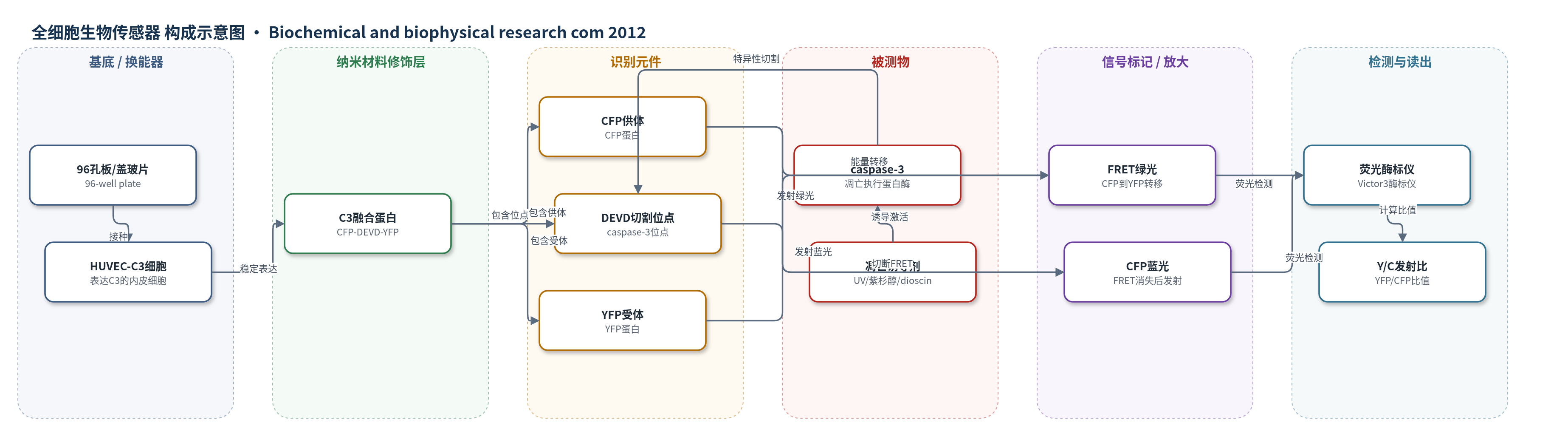
点击图片查看大图 · 依据论文自动绘制
传感器类型
全细胞生物传感器
检测对象
内皮细胞凋亡(endothelial cell apoptosis)、caspase-3 激活(caspase-3 activation);样品基质:HUVEC-C3 活细胞培养物(96孔板/盖玻片细胞培养体系)
检测原理
该传感器以稳定表达 C3 融合蛋白(CFP-DEVD-YFP)的 HUVEC-C3 细胞为全细胞传感平台。正常内皮细胞中,CFP 与 YFP 空间邻近,激发 CFP 后发生 FRET,YFP 绿光增强,Y/C 发射比高。凋亡诱导剂(UV、paclitaxel、dioscin 等)使细胞内 caspase-3 激活,特异性切割 DEVD 位点,导致 CFP 与 YFP 分离,FRET 被破坏,CFP 蓝光增强而 YFP 绿光减弱,Y/C 比下降。荧光酶标仪在 440 nm 激发下分别测量 486 nm CFP 和 535 nm YFP 发射,计算 Y/C 比,从而实时、定量反映 caspase-3 激活和凋亡程度。
检测灵敏度
CC50: paclitaxel FRET 0.71 ± 0.11 nM, MTT 1.20 ± 0.04 nM; vincristine FRET 2.29 ± 0.09 nM, MTT 3.06 ± 0.04 nM; dioscin MTT 2.18 ± 0.56 μM
效应效果
该体系能区分凋亡与坏死:H2O2(5 mM)和 Triton X-100(0.1%)使活力接近0,但Y/C比基本不变(6 h为5.37±0.77和6.28±0.41,对照5.75±0.63)。Z-VAD-FMK(40 μM)显著抑制paclitaxel诱导的Y/C下降,说明依赖caspase-3。Z0为0.66(>0.5)。与MTT相比更敏感:paclitaxel CC50为0.71±0.11 nM(MTT 1.20±0.04 nM),vincristine为2.29±0.09 nM(MTT 3.06±0.04 nM)。dioscin 5、10 μM降低Y/C,10 μM 24 h增加caspase-3活性并诱导PARP切割,CC50为2.18±0.56 μM,具VDA潜力。
传感器的构成
- 培养基底:96-well plate 或 coverslip/Petri dish,承载 HUVEC-C3 细胞并适配酶标仪或荧光显微镜。
- 传感细胞:HUVEC-C3 稳定细胞系,持续表达 C3 FRET 传感器,作为全细胞传感平台。
- 细胞培养介质:DMEM/F12、10% FBS 和 500 μg/mL geneticin,维持细胞存活与传感器稳定表达。
- 荧光供体:CFP(cyan fluorescent protein),接受激发后向 YFP 传递能量,切割后发射蓝光。
- 识别切割位点:DEVD(Asp-Glu-Val-Asp),caspase-3 凋亡激活后的特异性切割序列。
- 荧光受体:YFP(yellow fluorescent protein),接受 CFP 能量后发射绿光,用于计算 Y/C 比。
- 荧光读出:Zeiss Axiovert 100 荧光显微镜或 Perkin-Elmer Victor3 酶标仪,测量 CFP/YFP 发射强度。
中文摘要
本研究建立了一种基于荧光共振能量转移(FRET)的高通量内皮细胞凋亡检测方法。作者将 caspase-3 特异性 FRET 生物传感器 C3(CFP-DEVD-YFP)稳定转染人脐静脉内皮细胞(HUVEC),构建 HUVEC-C3 细胞系。正常状态下 CFP 激发后能量转移至 YFP,细胞呈绿色荧光;当紫外线或紫杉醇等凋亡诱导剂激活 caspase-3 并切割 DEVD 位点时,FRET 消失,荧光由绿转蓝。该方法在 96 孔板中通过测量 YFP/CFP 发射比实现高通量、实时监测,Z0 因子大于 0.5,表明适合高通量筛选。作者进一步用该体系筛选血管破坏剂(VDA),从内部化合物库中鉴定出 dioscin 为活性化合物。该检测可用于活细胞内皮凋亡监测和新 VDA 候选物发现。
英文摘要
In this study, we developed a high-throughput endothelial cell apoptosis assay using a fluorescence resonance energy transfer (FRET)-based biosensor. After exposure to apoptotic inducer UV-irradiation or anticancer drugs such as paclitaxel, the fluorescence of the cells changed from green to blue. We developed this method into a high-throughput assay in 96-well plates by measuring the emission ratio of yellow fluorescent protein (YFP) to cyan fluorescent protein (CFP) to monitor the activation of a key protease, caspase-3, during apoptosis. The Z' factor for this assay was above 0.5 which indicates that this assay is suitable for a high-throughput analysis. Finally, we applied this functional high-throughput assay for screening vascular disrupting agents (VDA) which could induce endothelial cell apoptosis from our in-house compounds library and dioscin was identified as a hit. As this assay allows real time and sensitive detection of cell apoptosis, it will be a useful tool for monitoring endothelial cell apoptosis in living cell situation and for identifying new VDA candidates via a high-throughput screening.