组成图示
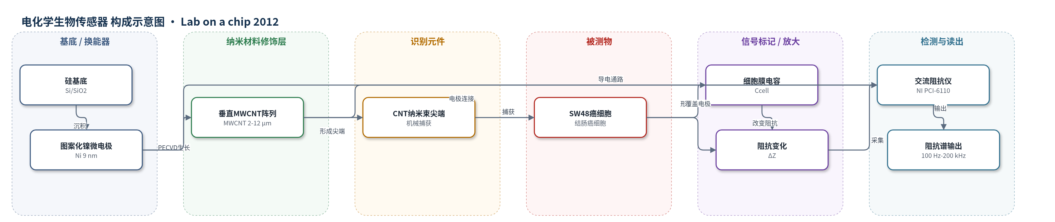
点击图片查看大图 · 依据论文自动绘制
传感器类型
电化学生物传感器
检测对象
SW48 结肠癌细胞(SW48 colon cancer cells),样品基质为 RPMI-1640 细胞悬液
检测原理
CNT-ECIS 以垂直排列 MWCNT 阵列同时作为粘附层和导电电极。当 SW48 癌细胞悬液流过电极表面时,癌细胞被弹性 MWCNT 尖端快速机械捕获,CNT 尖端接触甚至进入细胞膜,消除传统 ECIS 中细胞与电极之间的介质间隙。在交流探针信号下,细胞膜表现为电容 Ccell,CNT 阵列表现为低阻 RCNT,细胞间培养基间隙表现为 Rcell,溶液界面还包含 Helmholtz 双电层电容 CI 和扩散电阻 Rs。细胞覆盖使电极有效阻抗发生变化,细胞密度越高,阻抗变化越大。该过程无需聚合物粘附层、生物标志物或长时间贴壁,30 s 内即可完成捕获与信号读出,实现无标记、快速、高灵敏的癌细胞检测。
检测灵敏度
最低可检测细胞密度: 4000 cells cm^-2;灵敏度: 1.7 × 10^-3 Ω cm^2;TEF: 3.36 × 10^-3;CEF: 0.425 × 10^-6
效应效果
LDH 实验表明癌细胞在 30 s 和 5 min 后仍存活,阻抗信号来自活细胞。SEM 显示无 CNT 区域未捕获细胞,说明信号主要来自 CNT 捕获的癌细胞。与其他阻抗细胞传感器相比,CNT-ECIS 在 4000 cells cm^-2、<30 s 内获得 1.7×10^-3 Ω cm^2 灵敏度。TEF 为 3.36×10^-3,高于其他最佳值 0.35×10^-3;CEF 为 0.425×10^-6,高于其他最高值 0.17×10^-7,分辨率提高一个数量级以上。器件可实时监测凋亡:细胞空气中放置 10 min 后阻抗下降。较细 CNT(r<30 nm)比粗 CNT(r<70 nm)阻抗更高、捕获细胞更多。
传感器的构成
- 基底/绝缘层:Si 基底与热生长 SiO2 层,提供机械支撑与绝缘
- 换能器电极:图案化 Ni 微电极(9 nm),作为 CNT 生长催化剂和导电电极
- 纳米材料修饰层:垂直排列 MWCNT 阵列(长度 2–12 μm,直径 20–75 nm),兼作粘附层与导电层
- 细胞捕获/识别元件:弹性 MWCNT 纳米束尖端,机械捕获 SW48 癌细胞并接触细胞膜
- 被测物:SW48 结肠癌细胞,细胞膜形成电容 Ccell
- 信号读出:交流阻抗谱(AC impedance),NI PCI-6110 数据采集卡,100 Hz–200 kHz,0.2 V 偏置
中文摘要
本文首次报道了一种基于垂直排列碳纳米管的电细胞阻抗传感生物传感器(CNT-ECIS),用于快速、高灵敏且特异性地检测癌细胞。该传感器利用癌细胞在垂直碳纳米管阵列上的快速捕获,使碳纳米管尖端与细胞膜发生机械和电学相互作用,从而改变传感器阻抗。器件通过光刻工艺在 Ni/SiO2/Si 层上制备,碳纳米管阵列由直流等离子体增强化学气相沉积(DC-PECVD)在 9 nm 厚图案化镍微电极上生长。SW48 结肠癌细胞悬液流过电极表面后,被弹性碳纳米管束捕获;碳纳米管阵列同时充当粘附层和导电层,阻抗变化在 30 s 内即可出现。CNT-ECIS 可检测低至 4000 cells cm−2 的癌细胞,灵敏度为 1.7×10⁻³ Ω cm2。作者定义时间效率因子(TEF)和细胞效率因子(CEF),其值显著高于其他细胞基电学生物传感器。
英文摘要
A novel vertically aligned carbon nanotube based electrical cell impedance sensing biosensor (CNT-ECIS) was demonstrated for the first time as a more rapid, sensitive and specific device for the detection of cancer cells. This biosensor is based on the fast entrapment of cancer cells on vertically aligned carbon nanotube arrays and leads to mechanical and electrical interactions between CNT tips and entrapped cell membranes, changing the impedance of the biosensor. CNT-ECIS was fabricated through a photolithography process on Ni/SiO(2)/Si layers. Carbon nanotube arrays have been grown on 9 nm thick patterned Ni microelectrodes by DC-PECVD. SW48 colon cancer cells were passed over the surface of CNT covered electrodes to be specifically entrapped on elastic nanotube beams. CNT arrays act as both adhesive and conductive agents and impedance changes occurred as fast as 30 s (for whole entrapment and signaling processes). CNT-ECIS detected the cancer cells with the concentration as low as 4000 cells cm(-2) on its surface and a sensitivity of 1.7 × 10(-3)Ω cm(2). Time and cell efficiency factor (TEF and CEF) parameters were defined which describe the sensor's rapidness and resolution, respectively. TEF and CEF of CNT-ECIS were much higher than other cell based electrical biosensors which are compared in this paper.