组成图示
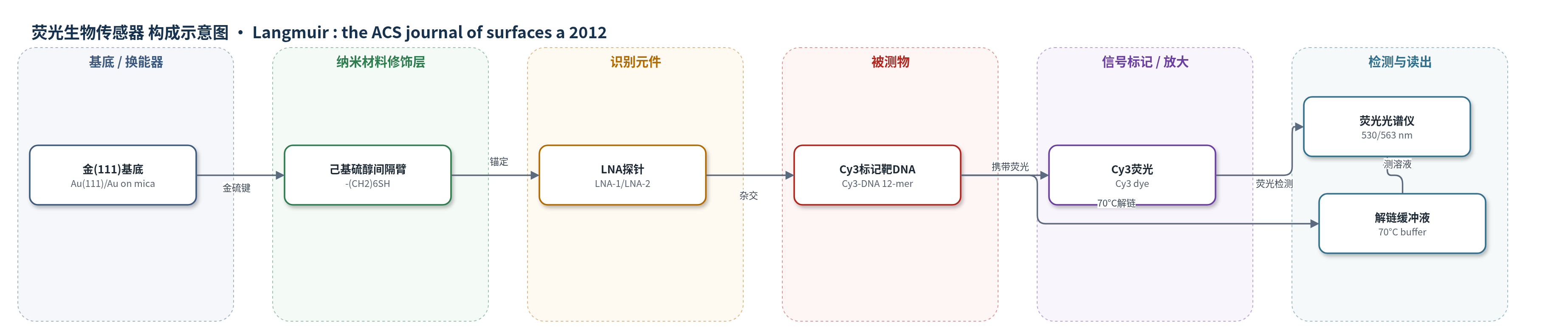
点击图片查看大图 · 依据论文自动绘制
传感器类型
荧光生物传感器
检测对象
互补单链 DNA 靶标(complementary ssDNA target,Cy3-DNA)、单碱基错配 DNA(single-base mismatch DNA);样品基质:磷酸盐缓冲液(20 mM 磷酸钠,100 mM NaCl,pH 7.0)
检测原理
硫醇化 LNA 探针通过 5′端己基硫醇链在金(111)表面形成 Au–S 键,己基间隔臂使 LNA 远离表面并逐步直立,减少碱基与金面的非特异吸附。Cy3 标记的靶 DNA 加入后,与表面 LNA 探针按 Watson–Crick 规则杂交;完全互补序列形成更稳定的 LNA–DNA 双链,单碱基错配序列稳定性较低。随后用 70°C 磷酸盐缓冲液解链,将已杂交的 Cy3-DNA 洗脱到溶液中,其荧光强度反映表面结合量。靶标结合越多,解链液中 Cy3 荧光越强。该体系无酶促或链式放大,主要依靠 LNA 的高亲和力、刚性骨架和有序直立单分子层提高识别保真度与信号强度。
检测灵敏度
原文未报告 LOD、线性范围、灵敏度斜率或相关系数。
效应效果
在相同条件下,LNA 自组装单分子层的绝对荧光信号约为 DNA 单分子层的 4–4.5 倍。完全匹配与单碱基错配的荧光强度比在 LNA 体系中约为 1.5,在 DNA 体系中约为 1.2;完全匹配相对单错配的荧光增强在 LNA 中约 50%,在 DNA 中约 26%,表明 LNA 的单碱基错配判别能力增强约 2 倍。AFM 显示 0.1 μM LNA 孵育 4 h 可形成 100–300 nm 长的一维 LNA ribbon,并平行排列成 7 μm × 7 μm 的二维有序膜;RAIR 表明 LNA 在数小时内由平躺转向直立。作者认为该有序 LNA 膜可用于表面核酸传感和单核苷酸多态性识别。
传感器的构成
- 基底:金(111)单晶表面(Au(111),金/云母,200 nm Au,火焰退火),提供 Au–S 化学吸附位点
- 间隔臂:5′端己基硫醇链(–(CH2)6SH),通过金–硫键锚定并抬高 LNA 以减少表面非特异吸附
- 识别元件:硫醇化锁核酸探针(ssLNA,LNA-1/LNA-2/LNA-3,12-mer),与互补 DNA 杂交识别
- 信号标记物:Cy3 荧光标记单链 DNA(Cy3-DNA,12-mer),作为荧光报告分子
- 缓冲介质:磷酸盐缓冲液(20 mM 磷酸钠,100 mM NaCl,pH 7.0),用于自组装、杂交、洗涤和解链
中文摘要
锁核酸(LNA)是一种构象受限的核酸类似物,因具有高效、序列特异性的 DNA/RNA 识别能力,且与固体表面相互作用弱于 DNA,有望成为核酸生物传感器中 DNA 的更优替代物。本文首次报道了一种基于简单浸渍法在金(111)表面构建有序、具有生物活性的 LNA 自组装单分子层的直接方法。该 LNA 层可产生比相应 DNA 层强 4–4.5 倍的 DNA 识别信号,并能区分完全互补 DNA 靶标与含单碱基错配的 DNA 靶标;其错配判别比约为 DNA 检测体系的 2 倍。高分辨原子力显微镜显示,LNA 分子形成数百纳米长的一维有序排列,并通过平行排布在大面积(7 μm × 7 μm)上形成二维有序组装。研究考察了 LNA 浓度和孵育时间对自组装的影响,并首次利用反射吸收红外光谱表征表面固定 LNA 的取向,发现 LNA 分子在数小时内由“平躺”构型转变为“直立”构型。
英文摘要
Locked nucleic acid (LNA) is a conformationally restricted nucleic acid analogue, which is potentially a better alternative than DNA for application in the nucleic acid based biosensor technologies, due to its efficient and sequence-specific DNA/RNA detection capability and lack of molecule-surface interaction on solid surfaces, compared to DNA. We report, for the first time, a straightforward way (based on simple immersion method) of generating an ordered self-assembled LNA monolayer, which is bioactive, onto a gold(111) surface. This layer is capable of giving rise to a stronger DNA recognition signal (4-4.5 times) than its DNA counterpart, and importantly, it can differentiate between a fully complementary DNA target and that having a single base mismatch, where the mismatch discrimination ratio is almost two times compared to the ratio relevant in case of DNA-based detection. We have presented high-resolution atomic force microscopy (AFM) topographs of the well-defined one-dimensional LNA molecular ordering (few hundred nanometers long) and of the two-dimensional ordered assembly formed over a large area (7 μm × 7 μm) due to parallel positioning of the one-dimensional ordered arrangements. The effects of different parameters such as LNA concentration and incubation time on LNA self-assembly have been investigated. Further, reflection absorption infrared (RAIR) spectroscopy has been applied to obtain information about the orientation of the surface-immobilized LNA molecules for the first time. It has been found that the LNA molecules undergo an orientational transition from the "lying down" to the "upright" configuration in a time scale of few hours.