组成图示
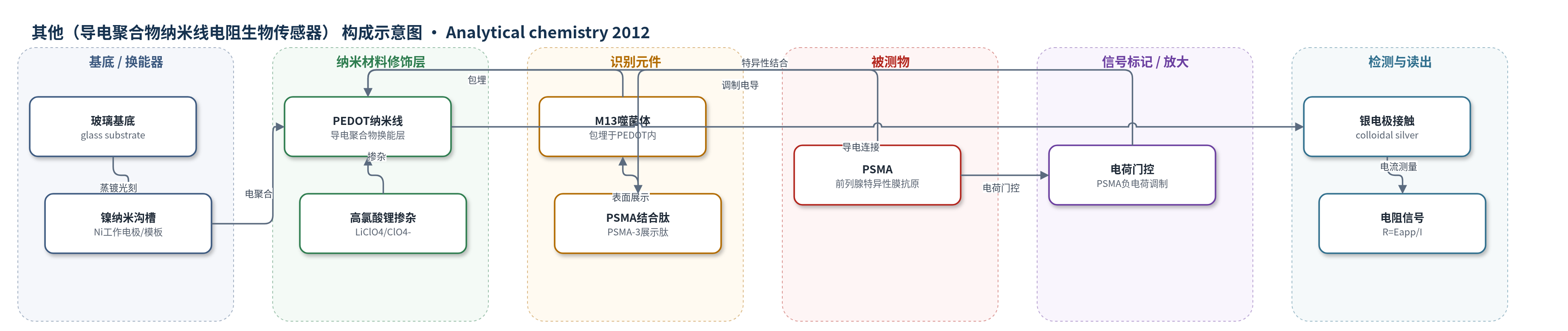
点击图片查看大图 · 依据论文自动绘制
传感器类型
其他(导电聚合物纳米线电阻生物传感器)
检测对象
前列腺特异性膜抗原(PSMA, prostate-specific membrane antigen);样品基质:高离子强度PBF缓冲液、合成尿(synthetic urine)
检测原理
传感器以PEDOT纳米线为导电换能层,M13噬菌体在电聚合时被包埋于PEDOT中,其表面展示的PSMA-3多肽作为识别元件。当PSMA进入PBF缓冲液或合成尿时,PSMA(pI约4.0,在pH 7.2下带负电)与展示肽特异性结合。结合事件使纳米线界面电荷分布改变,作者认为PEDOT矩阵相当于“分布式栅极”,PSMA的负电荷对PEDOT中的载流子进行门控,改变双极子/π电子的变程跳跃传输,使纳米线电阻增加。施加100 mV偏压并测量电流,由R=Eapp/I得到电阻变化;ΔR/R0随PSMA浓度在20–120 nM线性增大,从而实现无标记电阻检测。
检测灵敏度
LOD: 56 nM;线性范围: 20–120 nM;斜率: 2.1 × 10⁻³ nM−1;截距: 0.9 nM;回归残差标准差: 0.03
效应效果
该传感器对PSMA二聚体具有选择性,PSMA-3不结合PSMA单体、转铁蛋白受体(TfR)、酪蛋白及其他蛋白;阴性噬菌体无结合。抗干扰方面,器件可耐受干燥、洗涤、丙酮和硝酸处理,并能在150 mM盐的PBF缓冲液和合成尿中工作。实时检测中,PBF缓冲液LOD为56 nM,线性范围20–120 nM;合成尿中可测PSMA浓度高于66 nM。作者指出该灵敏度尚不足以检测血清PSMA,需提高2–3个数量级,但证明病毒-PEDOT纳米线可用于高离子强度体液中的癌症标志物检测。
传感器的构成
- 基底/模板:玻璃基底(glass substrate)上蒸镀镍膜(Ni),经光刻形成镍边缘纳米沟槽,作为工作电极与纳米线模板
- 导电聚合物换能层:PEDOT(poly(3,4-ethylenedioxythiophene))纳米线,由EDOT在LiClO4水溶液中电聚合形成,提供导电通路
- 掺杂/抗衡离子:LiClO4(高氯酸锂)提供ClO4-,与PEDOT阳离子结合并维持导电性
- 识别元件:M13噬菌体(M13 phage)颗粒,在电聚合中被包埋于PEDOT纳米线内
- 识别配体:PSMA-3噬菌体展示肽(PSMA-3 peptide,序列SECVEVFQNSCDW),特异性结合PSMA二聚体
- 电接触:胶体银(colloidal silver)涂覆形成两个电极接触,间隔约100 μm,用于接入电路
- 绝缘保护层:绝缘漆(insulating lacquer)覆盖银接触,防止银溶出
中文摘要
本文报道了一种基于含病毒聚(3,4-乙烯二氧噻吩)(PEDOT)纳米线从头构建的生物传感器,用于检测前列腺特异性膜抗原(PSMA)。该过程分三步:首先从约10^11个噬菌体展示肽库中筛选出表面展示多肽受体的M13噬菌体,该受体对PSMA具有高亲和力和选择性;其次采用光刻图案化纳米线电沉积(LPNE)方法微加工PEDOT纳米线,并在电聚合过程中包埋上述病毒颗粒;最后利用病毒-PEDOT纳米线阵列的直流电阻变化,在高离子强度(150 mM盐)介质(包括合成尿)中检测PSMA。在高离子强度磷酸缓冲氟化物(PBF)缓冲液中,纳米线阵列电阻随PSMA浓度在20–120 nM范围内线性增加,PSMA检出限为56 nM。
英文摘要
We demonstrate the de novo fabrication of a biosensor, based upon virus-containing poly(3,4-ethylene-dioxythiophene) (PEDOT) nanowires, that detects prostate-specific membrane antigen (PSMA). This development process occurs in three phases: (1) isolation of a M13 virus with a displayed polypeptide receptor, from a library of ≈10(11) phage-displayed peptides, which binds PSMA with high affinity and selectivity, (2) microfabrication of PEDOT nanowires that entrain these virus particles using the lithographically patterned nanowire electrodeposition (LPNE) method, and (3) electrical detection of the PSMA in high ionic strength (150 mM salt) media, including synthetic urine, using an array of virus-PEDOT nanowires with the electrical resistance of these nanowires for transduction. The electrical resistance of an array of these nanowires increases linearly with the PSMA concentration from 20 to 120 nM in high ionic strength phosphate-buffered fluoride (PBF) buffer, yielding a limit of detection (LOD) for PSMA of 56 nM.