组成图示
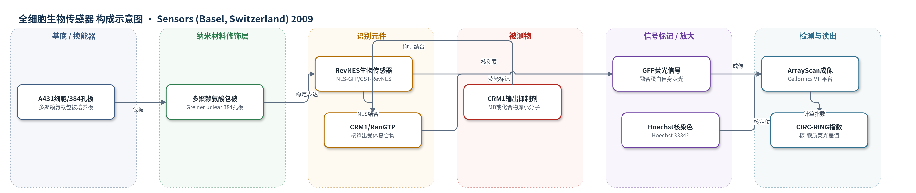
点击图片查看大图 · 依据论文自动绘制
传感器类型
全细胞生物传感器
检测对象
CRM1/Exportin1依赖核输出抑制剂(如LMB、ChemBioNet化合物库小分子);样品基质:活细胞培养体系(A431/HeLa等细胞,384孔板)
检测原理
该传感器以稳定表达NLS-GFP/GST-RevNES融合蛋白的活细胞为传感单元。融合蛋白含SV40 T抗原核定位信号(NLS)和HIV-1 Rev核输出信号(NES),正常状态下由CRM1/Exportin1与RanGTP介导核输出,稳态主要位于胞质。当CRM1依赖核输出抑制剂(如LMB或化合物库小分子)进入细胞并作用于CRM1、NES或相关共因子时,核输出受阻,融合蛋白在核内积累。Hoechst 33342标记细胞核,GFP荧光标记融合蛋白;ArrayScan VTI分别采集核CIRC区域和胞质RING区域荧光,计算CIRC-RING差值作为转移指数。抑制剂活性越强,核内GFP信号越高,CIRC-RING及Ti=compound(CIRC-RING)/DMSO(CIRC-RING)越大,从而实现小分子核输出抑制活性的定量检测。
检测灵敏度
原文未报告LOD、线性范围、灵敏度斜率或R^2。
效应效果
检测在384孔板中完成,每孔约470个有效细胞,扫描11 s/孔,48块板总扫描56 h,5天内完成16,671个化合物初筛。CIRC-RING的Z'因子为0.77,CIRCGFP为0.71,信噪比良好。初筛约500个潜在命中,经图像复查排除自荧光和细胞毒性假阳性后,120个化合物进入复筛,11个代表化合物经荧光显微镜验证;20个低于25 μM有效的化合物在多种细胞系中确认。生物传感器表达在选择培养基中至少稳定4个月。LMB处理5 h无明显细胞毒性,24 h降低活力;0.4% DMSO无检测效应。动力学监测可区分快/慢及可逆/不可逆抑制剂,作者认为可用于癌症化学基因组学。
传感器的构成
- 细胞基底:A431上皮癌细胞(A431bio),稳定表达RevNES-biosensor,提供活细胞传感平台。
- 培养/包被层:多聚赖氨酸(poly-L-lysine)包被的384孔薄底Greiner µclear板,用于细胞贴壁与成像。
- 识别/传感元件:NLS-GFP/GST-RevNES融合蛋白(RevNES-biosensor),含SV40 T抗原NLS与HIV-1 Rev NES,介导核质穿梭。
- 细胞内转运元件:CRM1/Exportin1与RanGTP,识别NES并介导核输出,是抑制剂作用靶点。
- 信号标记物:GFP荧光蛋白,作为融合蛋白自身荧光标记,无需外源染料。
- 核定位参考:Hoechst 33342核酸染料,标记细胞核,用于生成CIRC与RING掩膜。
- 换能/读出层:Cellomics ArrayScan VTI荧光显微成像平台,采集Hoechst与GFP通道并计算CIRC-RING转移指数。
中文摘要
荧光蛋白生物传感器可高时空分辨率解析细胞过程。受调控核质运输对多种生理病理反应至关重要。与遗传学方法不同,本研究建立并应用高内涵细胞转移生物传感器,用于化学基因组学解析核输出。通过抗生素筛选和流式分选,构建稳定表达GST-GFP及核输入/输出信号组合的A431细胞系。利用优化核转移算法,在Cellomics ArrayScan VTI平台稳健量化转移响应,并发展为384孔高内涵检测,筛选17K ChemBioNet化合物库。自动多参数分析结合图像检查,可区分真正核输出抑制剂与自荧光或细胞毒性假阳性。120个潜在命中进入复筛,20个低于25 μM有效的化合物在多种细胞系中确认。动力学分析识别出可逆或不可逆干扰CRM1核输出的抑制剂。该策略可鉴定用于解析活细胞核质运输的化学基因组学工具。
英文摘要
Fluorescent protein biosensors are powerful cellular systems biology tools for dissecting the complexity of cellular processes with high spatial and temporal resolution. As regulated nucleo-cytoplasmic transport is crucial for the modulation of numerous (patho)physiological cellular responses, a detailed understanding of its molecular mechanism would open up novel options for a rational manipulation of the cell. In contrast to genetic approaches, we here established and employed high-content cellular translocation biosensors applicable for dissecting nuclear export by chemicogenomics. A431 cell lines, stably expressing a translocation biosensor composed of glutathione S-transferase, GFP and a rational combination of nuclear import and export signals, were engineered by antibiotic selection and flow cytometry sorting. Using an optimized nuclear translocation algorithm, the translocation response could be robustly quantified on the Cellomics Arrayscan(®) VTI platform. Subsequent to assay optimization, the assay was developed into a higher density 384-well format high-content assay and employed for the screening of the 17K ChemBioNet compound collection. This library was selected on the basis of a genetic algorithm used to identify maximum common chemical substructures in a database of annotated bioactive molecules and hence, is well-placed in the chemical space covered by bioactive compounds. Automated multiparameter data analysis combined with visual inspection allowed us to identify and to rationally discriminate true export inhibitors from false positives, which included fluorescent compounds or cytotoxic substances that dramatically affected the cellular morphology. A total of 120 potential hit compounds were selected for Cellomics Arrayscan(®) VTI based rescreening. The export inhibitory activity of 20 compounds effective at concentrations < 25 μM were confirmed by fluorescence microscopy in several cell lines. Interestingly, kinetic analysis allowed the identification of inhibitors capable to interfere with the export receptor CRM1-mediated nuclear export not only in an irreversible, but also in a reversible fashion. In sum, exploitation of biosensor based screening allows the identification of chemicogenomic tools applicable for dissecting nucleo-cytoplasmic transport in living cells.