组成图示
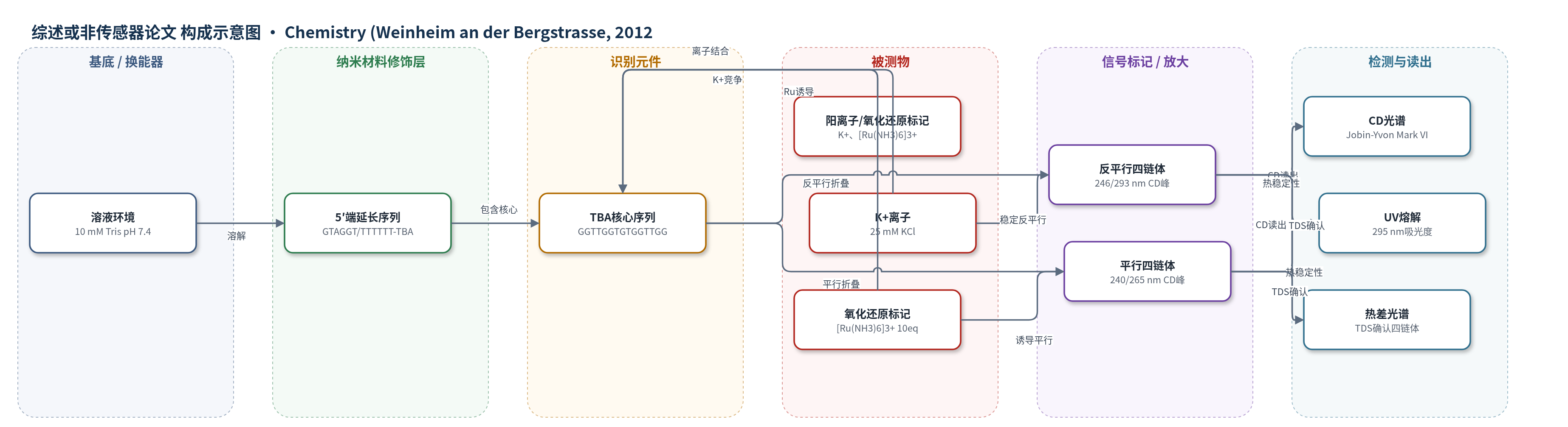
点击图片查看大图 · 依据论文自动绘制
传感器类型
综述或非传感器论文
检测对象
无实际检测对象;研究对象为血栓素结合适配体TBA及其5′端延长序列GTAGGT-TBA、TTTTTT-TBA的G-四链体构象;样品基质为10 mM Tris缓冲液(pH 7.4)及含K+、Na+、Mg2+、Ca2+、Sr2+、Ba2+、NH4+、Rb+或[Ru(NH3)6]2+/3+的溶液。
检测原理
本文并非构建完整传感器,而是研究适配体探针的离子敏感构象开关。TBA核心序列GGTTGGTGTGGTTGG在K+、Rb+、Sr2+、Ba2+等阳离子下形成反平行G-四链体,CD谱在约246 nm和293 nm出现正峰、267 nm附近出现负峰;5′端延长序列在Na+、Mg2+、Ca2+或[Ru(NH3)6]3+存在下可形成平行或平行/反平行混合结构,CD特征峰移至约240 nm负峰和260–275 nm正峰。[Ru(NH3)6]3+在每链10当量时诱导平行构象,加入K+后通过离子竞争使构象恢复反平行,呈现两态转换。UV熔解在295 nm监测超色效应,Tm反映结构稳定性;TDS进一步确认四链体特征。若用于电化学传感,构象变化可改变氧化还原标记与DNA磷酸骨架的可及性,从而产生可读出信号。
检测灵敏度
原文未报告LOD、线性范围、灵敏度斜率或相关系数。
效应效果
研究比较TBA、GTAGGT-TBA和TTTTTT-TBA在不同阳离子下的折叠与稳定性。TBA在所有研究阳离子下均保持反平行四链体;延长序列随阳离子呈现平行、反平行或混合结构。UV熔解Tm:TBA为Sr2+ 60°C、Ba2+ 57°C、K+ 45°C、Rb+ 29°C、NH4+ 28°C、Na+ 16°C;GTAGGT-TBA为Sr2+ 60°C、Ba2+ 55°C、K+ 35°C、Rb+ 18°C、NH4+ 20°C、Na+ 15°C;TTTTTT-TBA为Sr2+ 57°C、Ba2+ 52°C、K+ 33°C、Rb+ 15°C、NH4+ 14°C。热变性可逆且无滞后,提示分子内折叠。[Ru(NH3)6]3+与K+竞争实验显示两态构象开关,GTAGGT-TBA在0.5 mM K+下仍保持平行特征,而TTTTTT-TBA已转为反平行。作者认为该结果对TBA生物传感器设计重要,可避免阳离子诱导假阳性,并为电化学适配体传感器和氧化还原响应器件提供依据。
传感器的构成
- 识别元件:血栓素结合适配体TBA(5′-GGTTGGTGTGGTTGG-3′),作为识别thrombin的G-四链体核心序列。
- 延长探针:GTAGGT-TBA(5′-GTAGGTGGTTGGTGTGGTTGG-3′)和TTTTTT-TBA(5′-TTTTTTGGTTGGTGTGGTTGG-3′),5′端增加核苷酸以改变折叠与表面可及性。
- 阳离子/氧化还原标记:K+、Na+、Mg2+、Ca2+、Sr2+、Ba2+、NH4+、Rb+及[Ru(NH3)6]2+/3+,用于稳定不同四链体构象或作为电化学氧化还原探针。
- 溶液基质:10 mM Tris缓冲液(pH 7.4),提供结构研究缓冲环境。
中文摘要
基于适配体的生物传感器为蛋白的高性能、特异性检测提供了前景。血栓素结合适配体(TBA)是一种可形成G-四链体的DNA序列,在生物传感器构型中常在一端进行延长,以提高分析性能。本文研究了TBA在5′端延长对其结构和稳定性的影响。圆二色光谱表明,TBA在所有研究的阳离子(Ba2+、Ca2+、K+、Mg2+、Na+、NH4+、Sr2+以及[Ru(NH3)6]2+/3+氧化还原标记)存在下均折叠为反平行G-四链体构象;而延长适配体在部分阳离子存在下采取其他结构。基于紫外熔解曲线评估了各结构的稳定性,热差光谱确认所有构象均具有四链体特征。延长序列可根据阳离子性质采取平行或反平行结构,从而可能赋予离子敏感开关行为。该开关性质用常用氧化还原复合物[Ru(NH3)6]3+加以证明:其在极低浓度(每链10当量)下诱导平行构象;加入大量K+后构象恢复为反平行形式,为电化学生物传感或氧化还原响应器件提供了有趣前景。
英文摘要
Aptamer-based biosensors offer promising perspectives for high performance, specific detection of proteins. The thrombin binding aptamer (TBA) is a G-quadruplex-forming DNA sequence, which is frequently elongated at one end to increase its analytical performances in a biosensor configuration. Herein, we investigate how the elongation of TBA at its 5' end affects its structure and stability. Circular dichroism spectroscopy shows that TBA folds in an antiparallel G-quadruplex conformation with all studied cations (Ba(2+), Ca(2+), K(+), Mg(2+), Na(+), NH(4)(+), Sr(2+) and the [Ru(NH(3))(6)](2+/3+) redox marker) whereas other structures are adopted by the elongated aptamers in the presence of some of these cations. The stability of each structure is evaluated on the basis of UV spectroscopy melting curves. Thermal difference spectra confirm the quadruplex character of all conformations. The elongated sequences can adopt a parallel or an antiparallel structure, depending on the nature of the cation; this can potentially confer an ion-sensitive switch behavior. This switch property is demonstrated with the frequently employed redox complex [Ru(NH(3))(6)](3+), which induces the parallel conformation at very low concentrations (10 equiv per strand). The addition of large amounts of K(+) reverts the conformation to the antiparallel form, and opens interesting perspectives for electrochemical biosensing or redox-active responsive devices.