组成图示
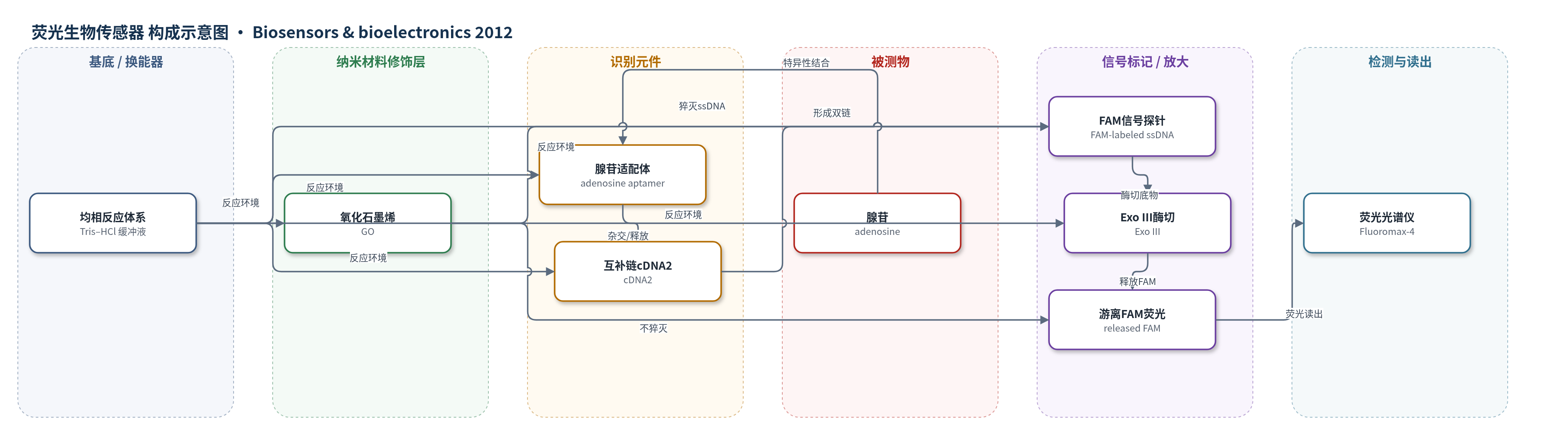
点击图片查看大图 · 依据论文自动绘制
传感器类型
荧光生物传感器
检测对象
腺苷(adenosine, Ado);样品基质:Tris–HCl 缓冲液/水溶液标准样品(未报道血清等实际生物基质)
检测原理
在无腺苷时,腺苷适配体与 cDNA2 杂交,信号探针保持单链;加入 GO 后,单链 FAM 探针通过 π-π 堆积被 GO 吸附并猝灭,荧光低。加入腺苷后,适配体优先结合腺苷,构象改变使 cDNA2 释放,cDNA2 与 FAM 信号探针杂交形成双链 DNA。Exo III 从信号探针 3′ 平末端/凹陷末端进行序列非特异性、过程性外切,循环切割双链并释放 5′-FAM 荧光基团;释放的 FAM 或极短片段不被 GO 有效猝灭,荧光增强。腺苷浓度越高,触发形成的双链越多,释放 FAM 越多,517 nm 荧光越强。
检测灵敏度
LOD: 1 nM;线性范围: 5 nM–100 μM(对数线性);回归方程: F = 1.30×10^5 log[Cadenosine] + 1.87×10^6;灵敏度斜率: 1.30×10^5(F 对 log[Cadenosine]);相关系数: 0.99
效应效果
该传感器对腺苷选择性良好:尿嘧啶、鸟嘌呤、胞嘧啶(200 μM)干扰下相对荧光响应仅约 -5.3%–7.0%;腺苷(100 μM)与三者(各 46.8 μM)混合时荧光仅轻微下降。检测限 1 nM,信号增强 >100%,低于常见荧光适配体传感器,并与近年报道相当。原文未报告稳定性、RSD和实际样品回收率。方法为均相检测,无需复杂电极修饰或分离步骤,作者认为其稳健、低成本、易自动化,可平行检测数百样品,适用于分子诊断、化学生物学和药物开发。
传感器的构成
- 反应介质:Tris–HCl 缓冲液(20 mM Tris,pH 8.0;腺苷标准液含 100 mM NaCl),提供杂交与酶切环境
- 识别元件:腺苷适配体(adenosine aptamer),特异性识别腺苷并触发杂交重排
- 互补链:cDNA2(GTTCCTCCGCAATACTTTTT),无靶标时结合适配体,有靶标时释放并与信号探针杂交
- 信号探针:5′-FAM 标记单链 DNA(FAM-TATTGCGGAGGAAC),携带荧光基团并被 Exo III 切割
- 信号放大酶:III 型外切酶(Exo III),从 3′ 末端循环切割双链 DNA,释放 FAM
- 猝灭/门控材料:氧化石墨烯(GO),通过 π-π 堆积吸附单链 FAM 探针并猝灭荧光
- 读出装置:荧光光谱仪(Fluoromax-4 spectrofluorometer),494 nm 激发、503–650 nm 发射检测
中文摘要
本文报道了一种基于氧化石墨烯(GO)的荧光适配体传感器,用于腺苷检测,并以III型外切酶(Exo III)作为信号放大元件。在无腺苷存在时,腺苷适配体与互补DNA(cDNA)杂交,Exo III不能切割5′端标记羧基荧光素(FAM)的单链信号探针;随后加入GO后,GO通过π-π堆积强烈吸附单链信号探针并有效猝灭荧光。当存在腺苷时,适配体优先结合靶标,使cDNA与信号探针形成双链DNA;Exo III随后从信号探针的3′平末端/凹陷末端消化双链DNA,释放FAM荧光基团。加入GO后,释放的FAM不能被有效吸附和猝灭。通过循环酶切放大,体系产生显著荧光增强。由于适配体对靶标的特异性识别以及GO对信号探针的强猝灭能力,该方法具有良好的选择性和高灵敏度。在优化条件下,信号增强超过100%,检测限低至1 nM,低于常用荧光适配体传感器。该传感器具有超高灵敏度,可作为临床诊断、分子生物学和药物开发的通用平台。
英文摘要
We report here a graphene oxide (GO)-based fluorescent aptasensor for adenosine detection by employing exonuclease III (Exo III) as a signal amplifying element. In the absence of adenosine, the adenosine aptamers hybridized with the complementary DNA (cDNA), and the Exo III could not cleave the single-strand signal probes labeled with carboxylfluorescein (FAM) at its 5' ends. When the graphene oxide was finally added, it could strongly adsorb the single-strand signal probes and quenched the fluorophore effectively. In the presence of adenosine, the aptamers associated with the targets, which led to the formation of duplex DNAs between the cDNAs and the signal probes. The Exo III thereafter could digest the duplex DNAs from 3' blunt terminus of signal probes, liberating the fluorophore. Upon adding the GO, the fluorophore could not be adsorbed and quenched. By coupling cyclic enzymatic cleavage, a remarkable fluorescent increase was obtained. Due to the specific recognition ability of the aptamer for the target and the powerful quenching property of GO for signal probe, this proposed approach has a good selectivity and high sensitivity for adenosine. In the optimum conditions described, >100% signal enhancement was achieved and a limit of detection as low as 1 nM was obtained, which is lower than those of commonly used fluorescent aptamer sensors. Moreover, the biosensor exhibited an ultrahigh sensitivity and held a versatile platform for clinical diagnostics, molecular biology and drug developments.