组成图示
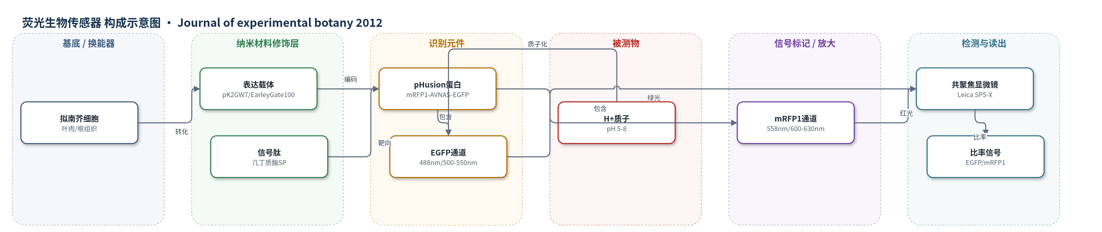
点击图片查看大图 · 依据论文自动绘制
传感器类型
荧光生物传感器
检测对象
pH(氢离子浓度,H+);样品基质:拟南芥叶肉细胞胞质、根组织质外体与胞质(活体植物组织)
检测原理
pHusion由pH敏感的EGFP与pH不敏感的mRFP1以1:1串联融合。当环境pH降低时,EGFP荧光团发生质子化,其荧光强度下降;mRFP1在生理pH范围内荧光基本不变,因此EGFP/mRFP1荧光比率随H+浓度升高而降低。传感器通过拟南芥几丁质酶信号肽或无信号肽分别定位到质外体/内膜系统或胞质,在活体组织中直接响应局部pH变化。共聚焦显微镜依次激发EGFP(488 nm)和mRFP1(558/585 nm),逐像素计算双通道比率,经Boltzmann曲线(pKa约6)换算为pH。该设计无需外源染料装载,可实现细胞与亚细胞水平的比率型活体pH成像。
检测灵敏度
未报告LOD、线性范围、灵敏度斜率或相关系数。
效应效果
pHusion在拟南芥全株稳定表达,未引起明显形态异常。体外共聚焦标定pKa约5.8,体内标定约6.0;外部pH在8至5.5间变化时,完整细胞胞质pH基本稳定(约6.4),而质外体pH快速可逆变化。医用胶固定会破坏根细胞质膜完整性,琼脂固定可使根保持活力数小时,13 min内无明显漂移。1 µM IAA处理使根伸长区皮下质外体立即碱化,估计ΔpH为0.5–0.8(pH 6–7范围),与对照相比P<0.001;成熟区无显著变化。灌注交换在1–3 s内完成,证明该传感器适合完整植物组织的动态pH生理研究。
传感器的构成
- 基底/宿主:拟南芥细胞(叶肉/根组织),作为传感器表达与活体成像基质。
- 表达载体:pK2GW7/pEarleyGate100,35S组成型启动子驱动pHusion表达。
- 定位元件:拟南芥几丁质酶信号肽(chitinase signal peptide),用于apo-pHusion靶向质外体。
- 传感蛋白:pHusion(mRFP1–AVNAS–EGFP),串联融合EGFP与mRFP1,实现比率型pH检测。
- pH响应元件:EGFP(enhanced green fluorescent protein),pKa约6.15,低pH下荧光淬灭,提供pH敏感信号。
- 内参荧光元件:mRFP1(monomeric red fluorescent protein),pKa约4.5,生理pH范围基本不敏感,作为比率参考。
- 读出换能器:Leica SP5-X共聚焦激光扫描显微镜,488 nm激发EGFP、558/585 nm激发mRFP1,双通道比率成像。
中文摘要
细胞内pH变化已被广泛认为是重要的信号机制。在植物中,质膜和液泡膜上的质子泵对维持胞内pH稳态及跨膜质子梯度起关键作用。为实时追踪外部刺激下pH在空间和时间上的精细变化,作者开发了一种新型遗传编码生物传感器pHusion。pHusion由增强型绿色荧光蛋白(EGFP)与单体红色荧光蛋白(mRFP1)串联融合而成,可作为比率型pH传感器,尤其适用于质外体pH测量。该传感器在拟南芥中组成型表达,并分别靶向胞质或质外体。由于活体显微成像高度依赖样品固定,而常用医用胶会破坏根细胞活力,作者建立了一种不损伤细胞活力的拟南芥幼苗根固定与灌注系统。利用该系统,作者成功追踪了叶肉细胞和根组织中质外体与胞质pH的变化:外部pH改变时,胞质pH波动很小,而质外体pH发生显著变化。外源吲哚-3-乙酸处理可立即引起根伸长区皮下质外体碱化,且该效应在伸长区显著强于根毛区及对照,展示了在完整组织生理背景下分析pH调控的潜力。
英文摘要
Changes in pH are now widely accepted as a signalling mechanism in cells. In plants, proton pumps in the plasma membrane and tonoplast play a key role in regulation of intracellular pH homeostasis and maintenance of transmembrane proton gradients. Proton transport in response to external stimuli can be expected to be finely regulated spatially and temporally. With the ambition to follow such changes live, a new genetically encoded sensor, pHusion, has been developed. pHusion is especially designed for apoplastic pH measurements. It was constitutively expressed in Arabidopsis and targeted for expression in either the cytosol or the apoplast including intracellular compartments. pHusion consists of the tandem concatenation of enhanced green fluorescent protein (EGFP) and monomeric red fluorescent protein (mRFP1), and works as a ratiometric pH sensor. Live microscopy at high spatial and temporal resolution is highly dependent on appropriate immobilization of the specimen for microscopy. Medical adhesive often used in such experiments destroys cell viability in roots. Here a novel system for immobilizing Arabidopsis seedling roots for perfusion experiments is presented which does not impair cell viability. With appropriate immobilization, it was possible to follow changes of the apoplastic and cytosolic pH in mesophyll and root tissue. Rapid pH homeostasis upon external pH changes was reflected by negligible cytosolic pH fluctuations, while the apoplastic pH changed drastically. The great potential for analysing pH regulation in a whole-tissue, physiological context is demonstrated by the immediate alkalinization of the subepidermal apoplast upon external indole-3-acetic acid administration. This change is highly significant in the elongation zone compared with the root hair zone and control roots.