组成图示
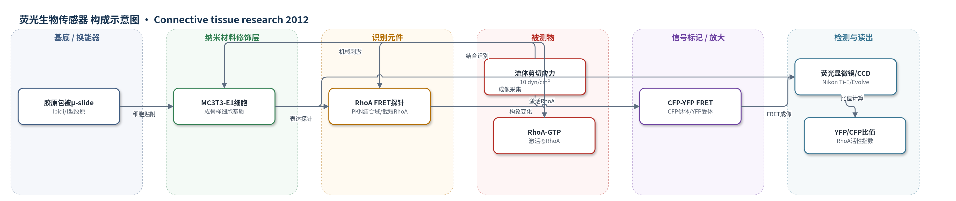
点击图片查看大图 · 依据论文自动绘制
传感器类型
荧光生物传感器
检测对象
RhoA激活状态(RhoA-GTP);样品基质:MC3T3-E1成骨样细胞(流体剪切处理)
检测原理
流体剪切应力作用于MC3T3-E1成骨样细胞后,细胞内小GTPase RhoA被激活并与GTP结合形成RhoA-GTP。RhoA-GTP与FRET探针中PKN的RhoA结合域结合,使截短RhoA构象改变,CFP供体与YFP受体距离缩短。CFP激发后,能量通过FRET转移至YFP,YFP发射增强,YFP/CFP发射比升高,从而反映RhoA激活水平。该过程不依赖酶催化沉积或核酸放大,信号放大主要来自FRET的距离依赖性和细胞内GTPase激活。通过倒置荧光显微镜分别采集CFP和YFP通道图像并计算比值,可实时监测机械刺激诱导的RhoA活性变化。
检测灵敏度
原文未报告LOD、线性范围、灵敏度斜率或相关系数。
效应效果
10 dyn/cm²流动处理1 h时,FRET探针显示RhoA约在40 min后激活;2–5 dyn/cm²未检测到Rho激活,提示应力阈值。C3转移酶降低基础RhoA活性并消除剪切诱导FRET变化,RhoA-N19共转染后无剪切诱导激活,说明特异性。Y27632在1 h时分别降低p-ERK1/2、p-p38和p-Akt磷酸化41%、58%和16%,3 h降低Cox2、Opn及Per2 mRNA,但不改变Per1和Per3。LY294002抑制PI3K后降低Opn表达。作者认为RhoA/ROCK位于PI3K和MAPK上游,是成骨细胞机械转导与骨形成调控的关键节点。
传感器的构成
- 基底/培养载体:I型胶原包被的μ-slide(Ibidi)或玻璃片,提供细胞贴附与流体剪切界面
- 宿主细胞层:MC3T3-E1成骨样细胞(经Neon电转表达FRET探针),作为感受机械刺激的细胞基质
- 识别元件:PKN的RhoA结合域(与截短RhoA偶联),结合RhoA-GTP并改变构象
- 信号标记物:CFP与YFP荧光蛋白突变体,通过FRET能量转移报告RhoA激活
- 光学换能/读出:Nikon Ti-E倒置显微镜、Evolve CCD与Semrock滤光片组,采集CFP/YFP发射比
中文摘要
成骨细胞在负载驱动骨形成中起关键作用,可通过骨细胞机械传感信号激活 Wnt 通路,但其对机械刺激的响应及小 GTPase RhoA 在机械转导中的作用尚不清楚。本研究以 MC3T3-E1 成骨样细胞为模型,在 10 dyn/cm² 单向流体剪切应力处理 1 h 条件下,检验 RhoA 信号是否介导细胞对流动剪切应力的响应。通过全基因组通路分析评估分子信号,并利用基于 CFP 与 YFP 荧光共振能量转移(FRET)的 RhoA 生物传感器测定 RhoA 激活状态。结果显示,流动处理激活磷脂酰肌醇 3-激酶(PI3K)和丝裂原活化蛋白激酶(MAPK)通路以及昼夜节律调控通路;Western blot 显示 Akt、p38 和 ERK1/2 磷酸化升高;FRET 测量表明 RhoA 被流动激活。Rho 激酶抑制剂显著降低流动诱导的 p38、ERK1/2 和 Akt 磷酸化,以及骨桥蛋白(Opn)和环氧合酶-2(Cox2)mRNA 水平。结果表明,RhoA 在成骨细胞机械转导中激活 PI3K 和 MAPK 信号并调节昼夜节律通路。
英文摘要
Osteoblasts play a pivotal role in load-driven bone formation by activating Wnt signaling through a signal from osteocytes as a mechanosensor. Osteoblasts are also sensitive to mechanical stimulation, but the role of RhoA, a small GTPase involved in the regulation of cytoskeleton adhesion complexes, in mechanotransduction of osteoblasts is not completely understood. Using MC3T3-E1 osteoblast-like cells under 1 hr flow treatment at 10 dyn/cm(2), we examined a hypothesis that RhoA signaling mediates the cellular responses to flow-induced shear stress. To test the hypothesis, we conducted genome-wide pathway analysis and evaluated the role of RhoA in molecular signaling. Activity of RhoA was determined with a RhoA biosensor, which determined the activation state of RhoA based on a fluorescence resonance energy transfer between CFP and YFP fluorophores. A pathway analysis indicated that flow treatment activated phosphoinositide 3-kinase (PI3K) and mitogen-activated protein kinase (MAPK) signaling as well as a circadian regulatory pathway. Western blot analysis revealed that in response to flow treatment phosphorylation of Akt in PI3K signaling and phosphorylation of p38 and ERK1/2 in MAPK signaling were induced. FRET measurement showed that RhoA was activated by flow treatment, and an inhibitor to a Rho kinase significantly reduced flow-induced phosphorylation of p38, ERK1/2, and Akt as well as flow-driven elevation of the mRNA levels of osteopontin and cyclooxygenase-2. Collectively, the result demonstrates that in response to 1 hr flow treatment to MC3T3-E1 cells at 10 dyn/cm(2), RhoA plays a critical role in activating PI3K and MAPK signaling as well as modulating the circadian regulatory pathway.