组成图示
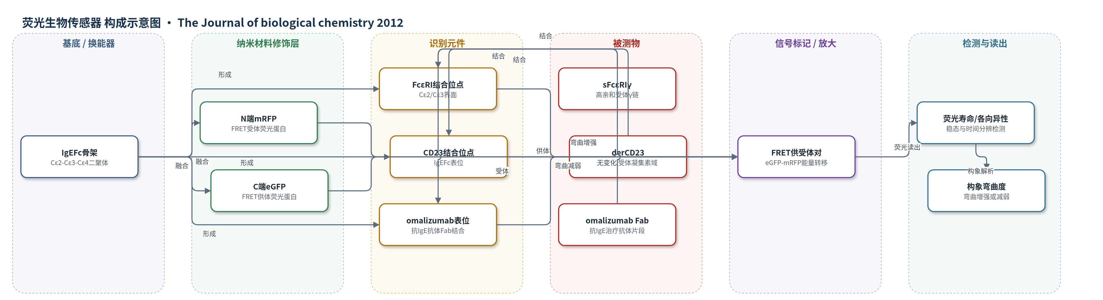
点击图片查看大图 · 依据论文自动绘制
传感器类型
荧光生物传感器
检测对象
人IgE Fc构象变化(human IgE Fc conformational change)、sFcεRIγ结合、derCD23结合、omalizumab Fab结合;样品基质:重组蛋白溶液/缓冲液(200 nM融合蛋白,20 °C)
检测原理
该传感器为溶液相FRET融合蛋白。eGFP作为供体被激发后,若与mRFP受体距离足够近且取向有利,则发生非辐射能量转移,FRET效率随供受体距离R的六次方变化,并受取向因子κ²影响。IgEFc的弯曲构象决定N端mRFP与C端eGFP的相对距离:游离IgEFc已呈弯曲,产生约9%的FRET;sFcεRIγ结合使IgEFc进一步弯曲,供受体距离缩短,FRET效率升高,eGFP寿命缩短、mRFP发射增强;omalizumab Fab结合使IgEFc伸展,距离增大,FRET降低,eGFP寿命延长;CD23结合不改变弯曲。HomoFRET各向异性进一步区分Cε2/Cε4域对内部运动与整体弯曲。
检测灵敏度
原文未报告 LOD、线性范围、灵敏度斜率或相关系数。
效应效果
融合荧光蛋白未明显改变IgEFc与sFcεRIγ的结合亲和力和动力学,SPR(Biacore)与野生型IgE无显著差异;DPI显示Cε2域使Fab/eGFP远离受体表面。稳态FRET效率为9.6±0.5%,寿命法为8.9±0.7%,与弯曲模型计算值9.2%一致,伸展模型仅0.54%。sFcεRIγ结合使eGFP激发加权平均寿命由2.45±0.01 ns降至2.38±0.01 ns;omalizumab Fab使其升至2.54±0.01 ns;derCD23无变化。HomoFRET各向异性在n=9重复中显示sFcεRIγ结合不改变Cε2/Cε4域对取向,支持刚性单元运动。作者认为该传感器验证晶体弯曲并非堆积假象,并解释受体结合IgE易被变应原交联。
传感器的构成
- 蛋白骨架/识别元件:人IgE Fc(IgEFc,Cε2-Cε3-Cε4二聚体),作为构象变化载体并携带sFcεRIγ、CD23和omalizumab结合位点
- 供体荧光标记:增强绿色荧光蛋白(eGFP),融合于IgEFc C端(双标mRFP-IgEFc-eGFP)或N端(eGFP-IgEFc对照),作为FRET供体
- 受体荧光标记:单体红色荧光蛋白(mRFP),融合于IgEFc N端,作为FRET受体
- 配体识别对象:可溶性FcεRI γ链(sFcεRIγ)、CD23凝集素域(derCD23)和omalizumab Fab,用于诱导或检测IgEFc构象变化
- 光学读出层:荧光光谱仪与时间分辨荧光寿命仪,检测eGFP/mRFP发射、寿命和各向异性
中文摘要
免疫球蛋白E(IgE)介导过敏反应,其Fc区与肥大细胞和嗜碱性粒细胞表面高亲和受体FcεRI的结合是过敏致敏与细胞激活的关键步骤。为在溶液中监测人IgE Fc(IgEFc)的构象及其受体结合时的变化,作者构建了基于Förster共振能量转移(FRET)的荧光生物传感器,将增强绿色荧光蛋白(eGFP)和单体红色荧光蛋白(mRFP)分别融合到IgEFc的N端和C端,并结合稳态激发扫描、荧光寿命和各向异性衰减进行定量。结果显示,游离IgEFc在溶液中呈弯曲构象;与可溶性FcεRI γ链(sFcεRIγ)结合后弯曲程度增强,而与低亲和受体CD23(FcεRII)结合无明显变化;抗IgE治疗抗体omalizumab的Fab结合则使IgEFc弯曲减弱。HomoFRET表明Cε2和Cε4域对在受体结合过程中作为刚性单元运动。该结果说明构象变化对FcεRI介导的IgE功能至关重要,并为受体结合IgE易被变应原交联提供了结构基础。
英文摘要
IgE binding to its high affinity receptor FcεRI on mast cells and basophils is a key step in the mechanism of allergic disease and a target for therapeutic intervention. Early indications that IgE adopts a bent structure in solution have been confirmed by recent x-ray crystallographic studies of IgEFc, which further showed that the bend, contrary to expectation, is enhanced in the crystal structure of the complex with receptor. To investigate the structure of IgEFc and its conformational changes that accompany receptor binding in solution, we created a Förster resonance energy transfer (FRET) biosensor using biologically encoded fluorescent proteins fused to the N- and C-terminal IgEFc domains (Cε2 and Cε4, respectively) together with the theoretical basis for quantitating its behavior. This revealed not only that the IgEFc exists in a bent conformation in solution but also that the bend is indeed enhanced upon FcεRI binding. No change in the degree of bending was seen upon binding to the B cell receptor for IgE, CD23 (FcεRII), but in contrast, binding of the anti-IgE therapeutic antibody omalizumab decreases the extent of the bend, implying a conformational change that opposes FcεRI engagement. HomoFRET measurements further revealed that the (Cε2)(2) and (Cε4)(2) domain pairs behave as rigid units flanking the conformational change in the Cε3 domains. Finally, modeling of the accessible conformations of the two Fab arms in FcεRI-bound IgE revealed a mutual exclusion not seen in IgG and Fab orientations relative to the membrane that may predispose receptor-bound IgE to cross-linking by allergens.