组成图示
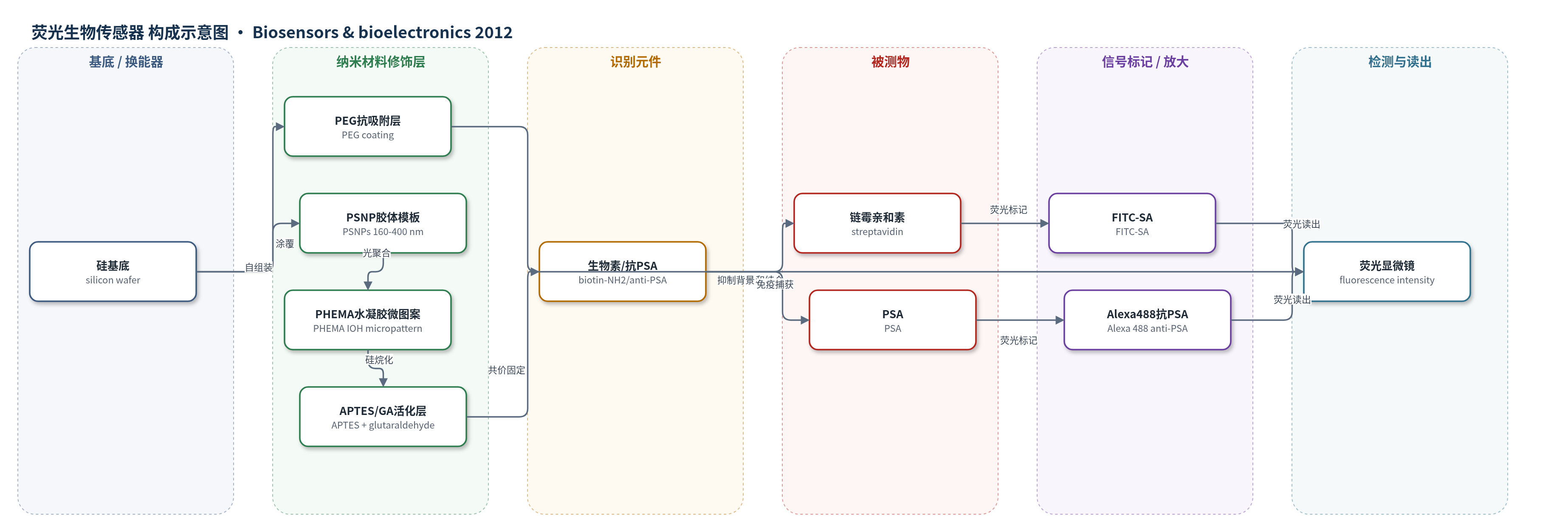
点击图片查看大图 · 依据论文自动绘制
传感器类型
荧光生物传感器
检测对象
链霉亲和素(streptavidin, SA,PBS 溶液)、前列腺特异性抗原(prostate-specific antigen, PSA,PBS 模拟血清/血清相关基质)
检测原理
该传感器基于三维反蛋白石水凝胶微图案上的特异性分子识别与荧光读出。PHEMA 水凝胶经 APTES 和戊二醛活化后,共价固定生物素或抗PSA抗体,形成高密度识别位点;PEG 背景抑制非特异吸附。当加入链霉亲和素时,FITC-SA 与固定生物素特异性结合;当检测 PSA 时,固定抗PSA先捕获 PSA,再加入 Alexa 488 标记抗PSA形成夹心复合物。识别事件使荧光标记物富集于微图案区域,荧光强度随目标物浓度增加而增强。反蛋白石大孔结构提供高内表面积和互连孔道,使蛋白负载和活性提高,相当于通过增加识别位点密度实现信号放大,最终由荧光显微镜测量荧光强度。
检测灵敏度
LOD: about 1.0 nM (IOH 240), 10.0 nM (IOH 400), 100 nM (non-porous);sensitivity: about 0.7 μM−1 (IOH 240), 0.46 μM−1 (IOH 400), 0.15 μM−1 (non-porous)
效应效果
PEG 背景有效抑制非特异吸附,链霉亲和素在 PEG 区非特异吸附不显著;各微图案荧光强度接近,表明位点均匀。BCA 显示 IOH240 固定蛋白 43.23±6.54 μg/cm2,IOH400 34.24±5.65 μg/cm2,非多孔 6.28±1.10 μg/cm2;未 APTES 修饰仅 1.30±0.35 和 0.97±0.24 μg/cm2,证明共价固定。GOX 活性随负载增加提高。水凝胶微图案在水中一周不脱落,长期应用建议丙烯酸酯硅烷锚定。生物素-链霉亲和素检测中 IOH240 LOD 1.0 nM、灵敏度 0.7 μM−1,优于 IOH400(10.0 nM、0.46 μM−1)和非多孔(100 nM、0.15 μM−1)。PSA 夹心免疫检测显示 IOH 荧光信号和灵敏度高于非多孔,作者认为可用于蛋白微阵列和多重生物传感。
传感器的构成
- 基底:硅片(silicon wafer),提供刚性支撑
- 抗吸附背景层:PEG 涂层(PEG coating),抑制非特异蛋白吸附
- 模板层:聚苯乙烯纳米颗粒(PSNPs)胶体晶体,自组装形成有序模板并去除后形成反蛋白石孔道
- 水凝胶微图案:PHEMA(聚(2-羟乙基甲基丙烯酸酯))水凝胶,由 HEMA、EGDMA 和 HOMPP 光聚合形成,提供三维大孔固定基质
- 化学修饰层:APTES(3-氨丙基三乙氧基硅烷)与 PHEMA 羟基反应引入氨基,并用戊二醛(GA)活化形成醛基
- 识别元件:生物素-NH2(biotin-NH2)或抗PSA抗体(anti-PSA),共价固定于水凝胶微图案用于特异性捕获
- 信号标记物:FITC-链霉亲和素(FITC-SA)或 Alexa Fluor 488 标记抗PSA抗体(Alexa 488 anti-PSA),提供荧光信号
- 封闭剂:BSA(牛血清白蛋白),用于 PSA 免疫检测中封闭非特异位点
- 读出:荧光显微镜/荧光强度测量(fluorescence microscopy/fluorescence intensity)
中文摘要
蛋白质图案化在生物传感器、生物MEMS、组织工程、细胞生物学和蛋白质组学研究中具有重要作用。本文开发了一种简单有效的蛋白质图案化技术,利用大孔聚(2-羟乙基甲基丙烯酸酯)(PHEMA)水凝胶微图案作为三维模板固定蛋白质。通过结合胶体晶体模板法与光刻,在聚乙二醇(PEG)涂层硅基底上制备了具有反蛋白石结构的大孔水凝胶微图案。所得反蛋白石水凝胶(IOH)微图案利用PHEMA中的羟基与3-氨丙基三乙氧基硅烷(APTES)反应,实现蛋白质的共价固定。蛋白质仅选择性固定在水凝胶微图案区域,而PEG区域作为有效的抗蛋白吸附屏障。由于高度有序且互连的三维大孔结构和较大的内表面积,IOH微图案中的蛋白负载约为非多孔水凝胶微图案的六倍,从而提高了蛋白活性。通过改变胶体纳米颗粒尺寸可控制水凝胶孔隙率,较小纳米颗粒可获得更高蛋白负载和活性。为展示其在生物传感器系统中的潜在应用,作者将生物素微图案化于水凝胶上,并成功利用IOH微图案检测链霉亲和素特异性结合,与相应非多孔水凝胶微图案相比具有更好的荧光信号和灵敏度。
英文摘要
Protein pattern has played an important role in biosensors, bioMEMS, tissue engineering, fundamental studies of cell biology, and basic proteomics research. Here, we developed a straightforward and effective protein patterning technique using macroporous poly(2-hydroxyethyl methacrylate) (PHEMA) hydrogel micropatterns as a three-dimensional (3D) template for protein immobilization. Micropatterns of macroporous hydrogels with inverse opal structures were prepared on poly(ethylene glycol) (PEG)-coated silicon substrates by combining a colloidal crystal templating method with photopatterning. The resultant inverse opal hydrogel (IOH) micropatterns were modified with 3-aminopropyltriethoxysilane using the hydroxyl groups in PHEMA for the covalent immobilization of proteins. Proteins were selectively immobilized only on the hydrogel micropatterns, while the PEG regions served as an effective barrier to protein adsorption. Because of their highly ordered and interconnected 3D macroporous structures and large internal surface areas, protein loading in the IOH micropattern was about six times greater than that on a non-porous hydrogel micropattern, which consequently improved the protein activity. The porosity of the hydrogel micropatterns could be controlled using different sizes of colloidal nanoparticles, and using smaller nanoparticles produced hydrogel micropatterns with higher protein loading capacities and activities. To demonstrate the potential use of IOH micropatterns in biosensor systems, biotin was micropatterned on the hydrogels and the specific binding of streptavidin was successfully assayed using IOH micropatterns with better fluorescence signals and sensitivity than that of the corresponding non-porous hydrogel micropatterns.