组成图示
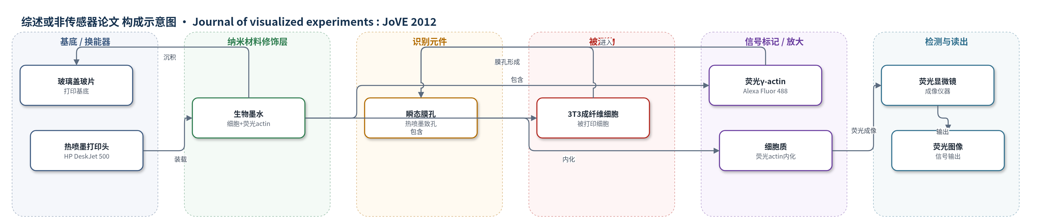
点击图片查看大图 · 依据论文自动绘制
传感器类型
综述或非传感器论文
检测对象
无明确检测对象;演示对象为3T3成纤维细胞(3T3 fibroblasts)与荧光标记γ-actin单体,样品基质为PBS细胞悬液/玻璃盖玻片。
检测原理
本文并非基于浓度响应的生物传感检测,而是利用热喷墨打印实现细胞内荧光标记物导入。含3T3成纤维细胞和荧光γ-actin单体的PBS生物墨水被装入改造后的HP DeskJet 500墨盒,打印头以热喷墨方式喷射约130 pL液滴到玻璃盖玻片上。液滴撞击与热喷墨过程可在细胞膜上形成约10 nm的瞬态孔,使原本难以跨膜的荧光actin单体进入细胞质;膜孔约2小时内自行关闭。荧光显微镜读取细胞内Alexa Fluor 488标记actin的荧光,用于观察细胞骨架动态。该过程不产生随被测物浓度变化的传感信号,因此不属于生物传感器。
检测灵敏度
原文未报告LOD、线性范围、灵敏度斜率或相关系数。
效应效果
作者将HP DeskJet 500改造为细胞打印平台,打印滴约130 pL,分辨率300 DPI。PBS比含血清培养基打印更一致且减少喷嘴堵塞;细胞在PBS中至少1小时仍保持活性,打印后活性与常规铺板相似。方法可在数分钟内处理数千个细胞,快于手动显微注射。荧光显微镜显示打印3 h后荧光actin分布于细胞质,15 min即可见细胞内荧光;DAPI核染色验证打印细胞,非生物材料不荧光。技术限制是仅适用于直径约10 nm以下分子或颗粒,且当前装置为二维打印。作者认为其可用于组织工程、基因转染、生物传感器微图案化和直接细胞治疗。
传感器的构成
- 非传感器说明:本文未构建生物传感器,仅报道热喷墨生物打印与细胞成像方法。
- 打印基底:玻璃显微镜盖玻片,承载细胞并用于荧光成像。
- 驱动/换能部件:HP DeskJet 500热喷墨打印头,产生约130 pL液滴并造成细胞膜瞬态孔。
- 生物墨水:3T3成纤维细胞与荧光标记γ-actin单体在PBS中的混合悬液,作为打印对象。
- 信号标记物:荧光标记γ-actin单体(Alexa Fluor 488),进入细胞后提供荧光信号。
- 读出方式:荧光显微镜,观察细胞内荧光actin分布。
中文摘要
生物打印在组织工程、直接细胞应用治疗和生物传感器微制造等方面具有广泛应用与重要意义。热喷墨打印也被用于基因转染,并显示可暂时破坏细胞膜而不影响细胞活性。膜上形成的瞬态孔可用于将通常无法穿过细胞膜的大分子引入细胞质。本文演示利用热喷墨打印将荧光标记的γ-肌动蛋白单体导入细胞。与显微注射相比,热喷墨打印对细胞相对温和,打印后细胞活性与常规铺板方法相似,并可在数分钟内处理数千个细胞。打印形成的孔约在2小时内关闭,但该技术形成的孔尺寸有限(约10 nm),因此仅适用于向细胞内注入小蛋白和/或颗粒。作者改造标准HP DeskJet 500打印机,使其可用于细胞打印:移除外壳、绕过进纸机构、制作载物台,并清洗墨盒。将3T3成纤维细胞与荧光γ-肌动蛋白单体混悬于PBS中,打印到玻璃盖玻片上,再用荧光显微镜成像,发现肌动蛋白分布于细胞质中。该导入方法可用于短时细胞骨架动态成像,具有多种应用价值。
英文摘要
Bioprinting has a wide range of applications and significance, including tissue engineering, direct cell application therapies, and biosensor microfabrication. Recently, thermal inkjet printing has also been used for gene transfection. The thermal inkjet printing process was shown to temporarily disrupt the cell membranes without affecting cell viability. The transient pores in the membrane can be used to introduce molecules, which would otherwise be too large to pass through the membrane, into the cell cytoplasm. The application being demonstrated here is the use of thermal inkjet printing for the incorporation of fluorescently labeled g-actin monomers into cells. The advantage of using thermal ink-jet printing to inject molecules into cells is that the technique is relatively benign to cells. Cell viability after printing has been shown to be similar to standard cell plating methods. In addition, inkjet printing can process thousands of cells in minutes, which is much faster than manual microinjection. The pores created by printing have been shown to close within about two hours. However, there is a limit to the size of the pore created (~10 nm) with this printing technique, which limits the technique to injecting cells with small proteins and/or particles. A standard HP DeskJet 500 printer was modified to allow for cell printing. The cover of the printer was removed and the paper feed mechanism was bypassed using a mechanical lever. A stage was created to allow for placement of microscope slides and coverslips directly under the print head. Ink cartridges were opened, the ink was removed and they were cleaned prior to use with cells. The printing pattern was created using standard drawing software, which then controlled the printer through a simple print command. 3T3 fibroblasts were grown to confluence, trypsinized, and then resuspended into phosphate buffered saline with soluble fluorescently labeled g-actin monomers. The cell suspension was pipetted into the ink cartridge and lines of cells were printed onto glass microscope cover slips. The live cells were imaged using fluorescence microscopy and actin was found throughout the cytoplasm. Incorporation of fluorescent actin into the cell allows for imaging of short-time cytoskeletal dynamics and is useful for a wide range of applications.