组成图示
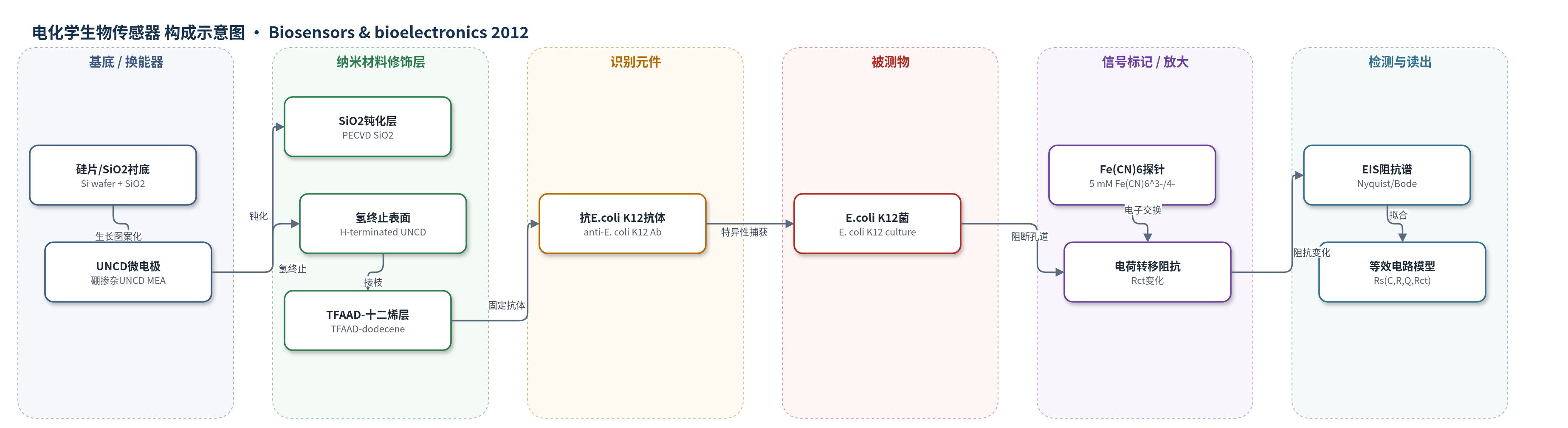
点击图片查看大图 · 依据论文自动绘制
传感器类型
电化学生物传感器
检测对象
大肠杆菌K12(Escherichia coli K12,E. coli K12);样品基质:热灭活E. coli K12菌悬液(约1.0×10^7 cfu/mL,PBS洗涤体系)
检测原理
该传感器采用无标记电化学阻抗谱(EIS)检测。抗大肠杆菌K12抗体固定于硼掺杂超纳米晶金刚石(UNCD)微电极表面,酪蛋白封闭非抗体区域,使电极表面形成由抗体位点构成的孔道。检测时,溶液中的Fe(CN)6^3-/4-氧化还原探针通过孔道与UNCD表面发生电子交换,其电荷转移过程表现为阻抗谱中的电荷转移电阻Rct。当E. coli K12与抗体特异性结合后,细菌体积阻塞孔道,降低有效电活性面积并减慢交换电流I0,使Rct增大;Rct2/Rct1比值反映捕获程度。微电极阵列增强传质并降低充电电流,提高信噪比。通过等效电路参数可区分特异结合与非特异结合。
检测灵敏度
灵敏度斜率: 微电极阵列 17 Ω ±10% per cfu/mL;短路阵列 0.03 Ω ±10% per cfu/mL;连续膜 0.005 Ω ±20% per cfu/mL;1000 cfu/mL时ΔRct=17 kΩ;灵敏度提高4个数量级。
效应效果
UNCD微电极阵列显著改善信号重现性,灵敏度较连续膜提高4个数量级。循环伏安峰电流在阵列微电极间变化<5%,>80%微电极可用。抗体/酪蛋白修饰后Rct为6.6–57 MΩ;捕获E. coli K12后,微电极3、4、8的Rct2/Rct1比值接近4.0,表明特异结合且非特异结合可忽略;微电极9出现Rct=103 MΩ、比值6.09,提示非特异结合。200 μm电极捕获50–100个细菌,10 μm电极约5–10个。1000 cfu/mL时Rct变化17 kΩ,与多数免疫分析检测限相当;作者认为可集成便携现场检测并扩展至多路复用。
传感器的构成
- 基底/换能器衬底:4英寸硅片(Si)及1 μm热氧化SiO2,提供机械支撑与绝缘衬底。
- 导电换能层:2 μm硼掺杂超纳米晶金刚石(UNCD)薄膜,光刻/刻蚀为3×3、200 μm微电极阵列(MEA),作为低背景电流阻抗换能电极。
- 钝化层:约1 μm PECVD SiO2,覆盖非电极区并隔离电解液,仅暴露微电极。
- 表面预处理:HFCVD原子氢环境氢终止(H-terminated)UNCD表面,提高生物分子光化学接枝效率。
- 固定化界面:TFAAD-十二烯单分子层(TFAAD-dodecene monolayer),用于抗体固定化并维持表面均匀性。
- 识别元件:抗大肠杆菌K12抗体(anti-E. coli K12 antibody,50 μg/mL),特异性捕获目标菌。
- 封闭层:酪蛋白封闭液(casein blocking solution),封闭非抗体位点,降低非特异结合。
- 电化学探针:5 mM K4Fe(CN)6/K3Fe(CN)6于0.01 M PBS(Fe(CN)6^3-/4-),作为EIS电子交换氧化还原探针。
中文摘要
无标记生物传感器能够实时检测抗原,并有望用于低成本、高特异、高灵敏的现场诊断与环境监测。基于电化学阻抗谱(EIS)的无标记生物传感器已用于细菌、病毒、DNA和蛋白质检测,但其商业化受到抗原结合后阻抗变化解释困难、表面污染和非特异结合导致信号重现性差等限制。本研究建立了一种电路模型,用于描述生物功能化表面的物理变化,并基于抗体–抗原周围孔道中电解液与电极表面之间的电子交换解释检测机制。该模型可定量提取不同功能化阶段生物表面的信息。此外,作者证明硼掺杂超纳米晶金刚石(UNCD)微电极阵列(3×3格式,200 μm直径)可显著提高信号重现性,并将灵敏度提高4个数量级。该工作首次展示了基于UNCD阵列的EIS生物传感器可靠检测模式大肠杆菌K12,为现场快速应用奠定基础。
英文摘要
It is well recognized that label-free biosensors are the only class of sensors that can rapidly detect antigens in real-time and provide remote environmental monitoring and point-of-care diagnosis that is low-cost, specific, and sensitive. Electrical impedance spectroscopy (EIS) based label-free biosensors have been used to detect a wide variety of antigens including bacteria, viruses, DNA, and proteins due to the simplicity of their detection technique. However, their commercial development has been hindered due to difficulty in interpreting the change in impedance upon antigen binding and poor signal reproducibility as a result of surface fouling and non-specific binding. In this study, we develop a circuit model to adequately describe the physical changes at bio functionalized surface and provide an understanding of the detection mechanism based on electron exchange between electrolyte and surface through pores surrounding antibody-antigen. The model was successfully applied to extract quantitative information about the bio surface at different stages of surface functionalization. Further, we demonstrate boron-doped ultrananocrystalline diamond (UNCD) microelectrode array (3 × 3 format, 200 μm diameter) improves signal reproducibility significantly and increases sensitivity by four orders of magnitude. This study marks the first demonstration of UNCD array based biosensor that can reliably detect a model Escherichia coli K12 bacterium using EIS, positioning this technology for rapid adoption in point-of-use applications.