组成图示
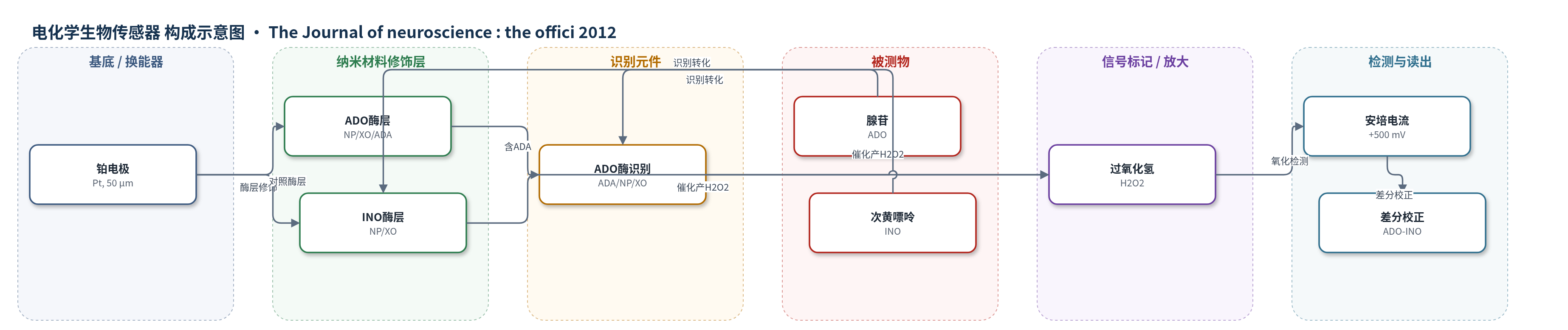
点击图片查看大图 · 依据论文自动绘制
传感器类型
电化学生物传感器
检测对象
腺苷(adenosine, ADO);次黄嘌呤(inosine, INO,对照背景);样品基质:小鼠海马急性切片细胞外环境(aCSF灌注)
检测原理
腺苷(ADO)进入ADO酶电极后,先被腺苷脱氨酶(ADA)转化为次黄嘌呤(INO),再由核苷磷酸化酶(NP)转化为黄嘌呤,黄嘌呤氧化酶(XO)催化其氧化为尿酸并生成过氧化氢(H2O2)。H2O2在铂电极+500 mV下发生安培氧化,产生与ADO浓度成正比的电流。INO对照电极不含ADA,仅响应INO及其下游嘌呤代谢物;实验将INO信号按标准灵敏度缩放后从ADO信号中扣除,得到ADO特异性电流,并用10 μM腺苷/次黄嘌呤标准校准。因此信号随细胞外ADO浓度升高而增大,酶级联提供化学转化而非额外信号放大。
检测灵敏度
线性范围: 0.33–10 μM;R = 0.99
效应效果
该酶电极采用ADO/INO成对差分策略,扣除次黄嘌呤和非特异电化学信号,提高腺苷选择性;10 μM腺苷/次黄嘌呤标准用于校准。ADO传感器在0.33–10 μM线性响应,R=0.99。作者估计灵敏度时变使绝对读数偏高约60%,但条件一致,不影响比较。海马切片中,WT觉醒和睡眠剥夺使细胞外腺苷升高,dnSNARE小鼠该升高减弱;睡眠剥夺dnSNARE组INO代谢物趋势升高(3.29±0.74 vs 1.51±0.56 μM,p=0.17)。作者主张该直接测量可弥补微透析采样比例有限和时间平均不足。
传感器的构成
- 换能器电极:铂电极(Pt electrode,50 μm直径),作为电化学换能器,在+500 mV下检测H2O2电流
- 酶修饰层(ADO传感器):核苷磷酸化酶(NP)、黄嘌呤氧化酶(XO)、腺苷脱氨酶(ADA),催化腺苷经次黄嘌呤、黄嘌呤转化为尿酸并产生H2O2
- 酶修饰层(INO对照传感器):NP、XO,不含ADA,对腺苷不敏感,用于检测次黄嘌呤/嘌呤代谢物背景
- 识别元件:ADA(ADO传感器中),赋予腺苷特异性;INO传感器以NP/XO识别次黄嘌呤
- 信号产物:过氧化氢(H2O2),由XO催化产生,被Pt电极安培氧化
- 校准介质:人工脑脊液(aCSF)及10 μM腺苷/次黄嘌呤标准品,用于水化、预校准和灵敏度校正
中文摘要
睡眠缺失导致睡眠驱动力增加和海马依赖记忆缺陷,这些反应被认为需要腺苷A1受体(adorA1R)激活以及星形胶质细胞释放ATP等递质,ATP在细胞外迅速转化为腺苷,即胶质传递。尽管星形胶质细胞来源腺苷在睡眠稳态和睡眠缺失影响记忆中作用日益明确,但此前研究未确定该信号分子浓度是否随觉醒升高。本文显示,小鼠中adorA1R激活水平随觉醒增加,并影响海马突触传递和皮层网络活动。基于生物传感器的直接测量表明,正常觉醒和睡眠剥夺使细胞外腺苷净浓度升高。遗传抑制胶质传递可阻止该升高,并减弱觉醒依赖的adorA1R介导突触与网络调节。因此,觉醒通过星形胶质细胞释放递质增加海马细胞外腺苷水平。
英文摘要
Loss of sleep causes an increase in sleep drive and deficits in hippocampal-dependent memory. Both of these responses are thought to require activation of adenosine A1 receptors (adorA1Rs) and release of transmitter molecules including ATP, which is rapidly converted to adenosine in the extracellular space, from astrocytes in a process termed gliotransmission. Although it is increasingly clear that astrocyte-derived adenosine plays an important role in driving the homeostatic sleep response and the effects of sleep loss on memory (Halassa et al., 2009; Florian et al., 2011), previous studies have not determined whether the concentration of this signaling molecule increases in response to wakefulness. Here, we show that the level of adorA1R activation increases in response to wakefulness in mice (Mus musculus). We found that this increase affected synaptic transmission in the hippocampus and modulated network activity in the cortex. Direct biosensor-based measurement of adenosine showed that the net extracellular concentration of this transmitter increased in response to normal wakefulness and sleep deprivation. Genetic inhibition of gliotransmission prevented this increase and attenuated the wakefulness-dependent changes in synaptic and network regulation by adorA1R. Consequently, we conclude that wakefulness increases the level of extracellular adenosine in the hippocampus and that this increase requires the release of transmitters from astroctyes.