组成图示
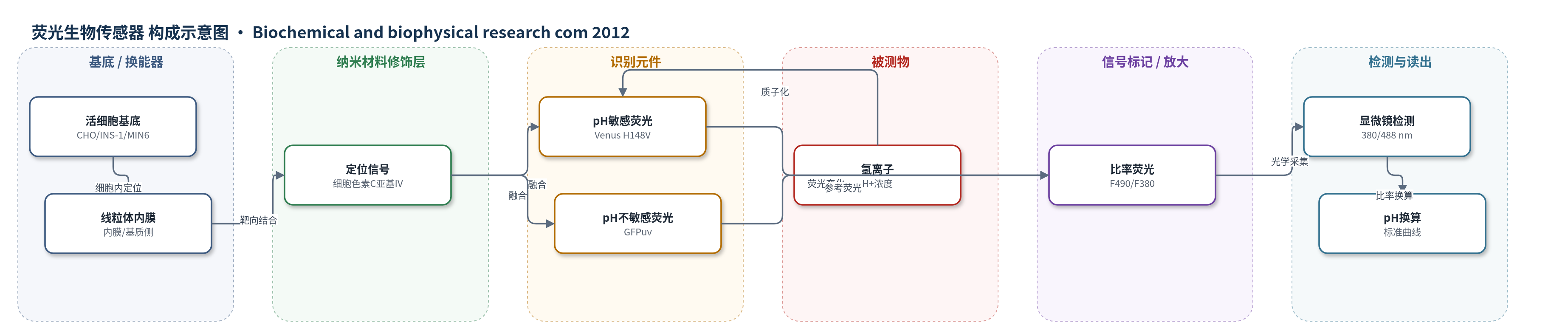
点击图片查看大图 · 依据论文自动绘制
传感器类型
荧光生物传感器
检测对象
线粒体pH(mitochondrial pH, pH_mito);样品基质:活细胞(CHO-K1、INS-1、MIN6胰腺β细胞系)线粒体内膜/基质侧
检测原理
MTpHGV由氢离子敏感的Venus H148V与氢离子不敏感的GFPuv融合,并经细胞色素C亚基IV信号靶向线粒体内膜。当线粒体内膜两侧H+浓度改变时,H+使Venus H148V敏感位点质子化,改变发色团质子化状态和荧光特性,使490 nm激发荧光强度随pH变化;GFPuv在380 nm激发下提供稳定参考。仪器同时采集双激发/单发射信号,计算F490/F380比率,消除表达量、细胞运动、光漂白和聚焦差异。CCCP、葡萄糖等改变线粒体代谢和H+梯度,引起比率变化,再按纯化探针pH 6.0–8.0标准曲线换算为线粒体pH。
检测灵敏度
未报告LOD、线性范围、灵敏度斜率或相关系数。
效应效果
该比率型探针氯离子不敏感、氢离子敏感,可避免表达量、细胞运动和光学聚焦造成的伪影,实现活细胞长时间无漂白监测。稳定转染细胞可培养约2个月。1 μM CCCP快速改变线粒体pH;葡萄糖刺激下,MTpHGV-INS-1细胞pH升高0.25±0.05(n=6),MTpHGV-MIN6细胞降低0.33±0.11(12–14细胞/皿,8皿/实验)。100 μM diazoxide消除Ca2+响应但不影响线粒体pH,5 μM AOA消除酸化,提示依赖NADH穿梭而非KATP通道。作者认为可用于评估活细胞线粒体功能及糖尿病β细胞代谢研究。
传感器的构成
- 基底/换能器:活细胞线粒体内膜与光学显微镜,提供传感微环境和荧光信号读出
- 传感元件:Venus荧光蛋白H148V突变体,氢离子敏感,提供pH依赖荧光变化
- 内参元件:GFPuv变体,氢离子不敏感,提供稳定参考荧光
- 融合探针:pHGV,由H148V-Venus与GFPuv融合,实现双激发/单发射或单激发/双发射比率测量
- 定位元件:细胞色素C亚基IV定位信号,将pHGV靶向线粒体内膜,形成MTpHGV
- 表达平台:CHO-K1、INS-1、MIN6细胞稳定转染MTpHGV,构建活细胞线粒体pH传感系统
- 体外校准:pGEX-4T融合探针纯化后进行pH 6.0–8.0滴定,建立荧光比率标准曲线
- 光学读出:共聚焦/倒置显微镜以380 nm与488/490 nm双激发、520 nm发射,计算F490/F380比率
- 辅助对照:MitoTracker Red标记线粒体,BCECF-AM测胞质pH,Fura-2AM测Ca2+
中文摘要
线粒体pH是线粒体能量代谢的关键决定因素。本研究开发了一种基于氯离子不敏感且氢离子敏感探针的新型荧光比率型pH生物传感器,用于活细胞中直接、定量且无光漂白地测量线粒体pH。该探针由氢离子敏感的Venus H148V突变体与氢离子不敏感的GFPuv融合而成,并融合线粒体定位信号形成MTpHGV。将MTpHGV稳定转染至大鼠INS-1和小鼠MIN6胰腺β细胞后,可连续监测线粒体pH。CCCP处理使INS-1细胞线粒体pH快速下降,而MIN6细胞快速上升,提示探针在INS-1中位于基质侧,在MIN6中位于线粒体内膜外侧。葡萄糖刺激使INS-1细胞pH快速升高,而MIN6细胞pH快速下降后恢复。在AOA或100 μM diazoxide条件下,葡萄糖诱导的线粒体酸化依赖NADH穿梭,而不依赖KATP通道。该体系在生理范围内敏感,可作为评估活细胞线粒体功能的工具。
英文摘要
Mitochondrial pH is a key determinant of mitochondrial energy metabolism. We have developed a new fluorescence-based ratiometric pH biosensor using a chloride-insensitive and hydrogen-sensitive probe for direct, quantitative and bleaching-free measurement in a living cell. Fusing this biosensor with a mitochondrial localization signal (MTpHGV) allowed us to determine mitochondrial pH. This new system was applied to measure mitochondrial pH in pancreatic β-cells, in which mitochondrial function plays a pivotal role in insulin secretion. Rat INS1 cells and mouse MIN6 cells are transfected with MTpHGV stably to monitor mitochondrial pH. While carbonyl cyanide 3-chlorophenylhydrazone (CCCP) treatment rapidly decreased mitochondrial pH in cultured rat MTpHGV-INS-1 cells, MTpHGV-MIN6 cells showed a rapid increase. These data suggest that MTpHGV probe exist in matrix side in INS-1 cells, but on the outer side of mitochondrial inner membrane in MIN6 cells. Moreover, while MTpHGV-INS-1 cells showed a rapid increase of pH by glucose stimulation, mitochondrial pH decreased quickly by glucose stimulation in all MTpHGV-MIN6 cells examined and recovered smoothly. Perfusion study of glucose load in MTpHGV-MIN6 cells under aminooxyacetate (AOA) or 100μM diazoxide showed that this mitochondrial pH acidification was dependent on nicotinamide adenine dinucleotide (NADH) shuttle, but independent from KATP channel. This new system for measuring mitochondrial pH is sensitive across the range of physiologic conditions and may be a useful tool for evaluating mitochondrial function in living cells.