组成图示
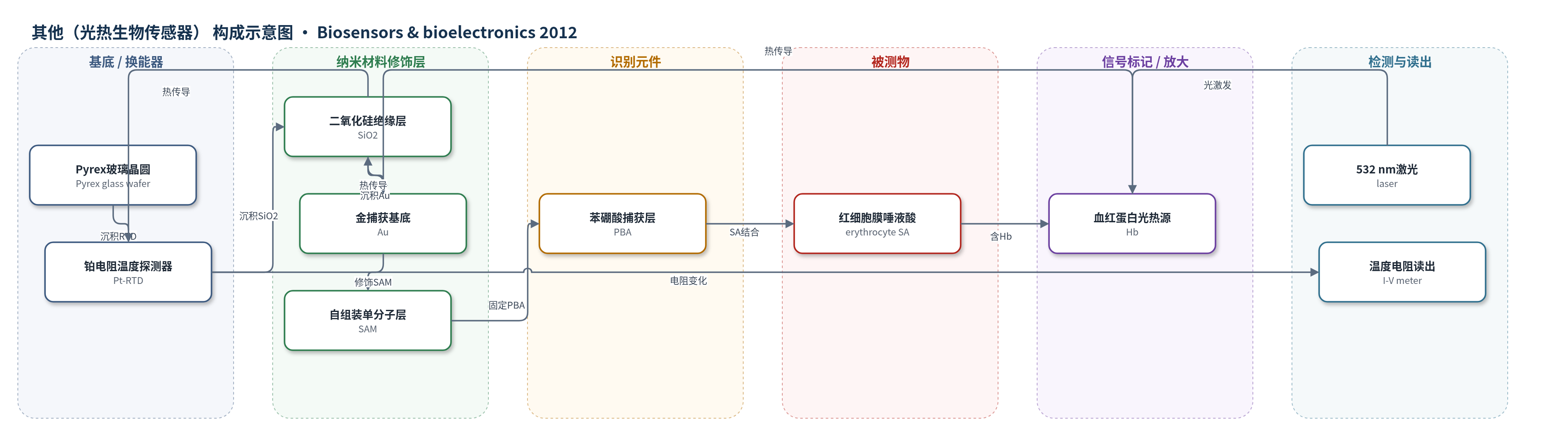
点击图片查看大图 · 依据论文自动绘制
传感器类型
其他(光热生物传感器)
检测对象
红细胞膜唾液酸(sialic acid, SA);样品基质:全血/洗涤红细胞(whole blood, washed erythrocytes)
检测原理
传感器表面PBA与红细胞膜SA发生可逆选择性结合,SA表达量越高,捕获红细胞越多。红细胞内血红蛋白(Hb)作为内源光热转换分子,吸收532 nm激光并转化为热。热通过SAM、金层和SiO2传导至底层Pt-RTD,使其电阻随温度升高而增加。Pt-RTD电阻与温度满足R(t)=1.8459t+601.34,通过I-V测量电阻并换算温度变化。温度变化与捕获红细胞数量及SA浓度成正比,利用红细胞内大量Hb实现内源信号放大,无需酶反应或外源标记。
检测灵敏度
LOD: 0.8 W/cm2 下 322.3 pmol/10^6 cells;1.6 W/cm2 下 246.6 pmol/10^6 cells;2.4 W/cm2 下 234.3 pmol/10^6 cells;3.2 W/cm2 下 219.7 pmol/10^6 cells
效应效果
系统可在约5 min内用1 μL全血完成检测,避免传统酶法>12 h、成本>$20/次及1–200 nmol窄范围限制,也避免FET的Debye长度限制。SA-PBA结合约50 s饱和,60 s最优;全血SA 379 pmol/10^6 cells捕获58±17 cells/mm2,ficin处理后捕获数随SA降低。0.01 M SA封闭后无温度变化和红细胞结合,显示特异性。RSD随激光功率升高总体降低:0.8 W/cm2为41.2%–91.9%,1.6 W/cm2为17.9%–209.9%,2.4 W/cm2为25.8%–54.6%,3.2 W/cm2为20.1%–63.4%。作者认为可用于糖尿病和癌症诊断。
传感器的构成
- 基底:Pyrex玻璃晶圆(Pyrex glass wafer),承载各功能层
- 换能器:铂电阻温度探测器(Pt-RTD, platinum resistive temperature detector),通过电阻变化测量温度
- 绝缘层:二氧化硅(SiO2),隔离上下层
- 捕获基底:金层(Au),承载SAM和PBA并参与光热吸收
- 修饰层:1-羧基-10-癸硫醇自组装单分子层(SAM, 1-carboxy-10-decanethiol),提供羧基固定PBA
- 识别元件:苯硼酸(PBA, phenylboronic acid),与红细胞膜唾液酸(SA)可逆选择性结合
- 信号标记物:红细胞内血红蛋白(Hb, hemoglobin),吸收532 nm激光产生热
中文摘要
红细胞膜唾液酸(SA)的定量分析是诊断癌症和糖尿病的重要临床参数,但现有方法数量少、成本高、耗时长且操作复杂。本文报道一种新型光热生物传感系统,用于直接测量暴露于红细胞膜表面的SA。该系统利用红细胞内血红蛋白吸收532 nm激光并转化为温度变化。首先,用含苯硼酸(PBA)的自组装单分子层(SAM)修饰微温度计传感器表面,以捕获表达SA的红细胞;随后充分洗涤,SA表达量越高,红细胞与传感器结合越紧密。最后,用底层微米级铂电阻温度探测器(Pt-RTD)测量经532 nm激光加热后血样的温度变化。捕获在传感器表面的红细胞所产生的温度变化可反映其膜上SA表达量。该方法避免了传统方法中的多重酶反应和长时间处理,有望为糖尿病和癌症诊断中SA表达水平的定量分析提供新工具。
英文摘要
The quantitative analysis of sialic acid (SA) at an erythrocyte membrane is becoming an important clinical parameter in diagnosing cancer and diabetes. In spite of such clinical importance, there are only a few, very expensive, time consuming and complicated quantifying methods established. To solve this problem, we demonstrate a novel and direct measurement technique for SA exposed to the cell membrane using a photothermal biosensing system in which the hemoglobin molecules in the erythrocyte absorb a specific wavelength of photons (532 nm) and convert it to a temperature change. For measuring the quantity of SA, we first modified the sensor surface of a micro-scaled thermometer using phenylboronic acid (PBA) containing a self-assembled monolayer (SAM) to capture the SA-expressing erythrocytes. Second, the sensor surface was thoroughly washed, and when more SA was expressed, tighter association of erythrocytes to the biosensor was expected. Thirdly, blood sample changes in temperature, heated by the 532 nm wavelength laser, were measured by the bottom layer's micron sized platinum thermometer. The temperature changes from the erythrocytes captured on the sensor surface could be estimated by the amount of SA expressed on the erythrocyte membrane. This novel SA analysis system can solve the problems raised by conventional methods such as multiple enzyme reactions and a time consuming process. We expect that this system will help provide a new tool in the quantitative analysis of SA expression level for the diagnosis of diabetes and cancers.