组成图示
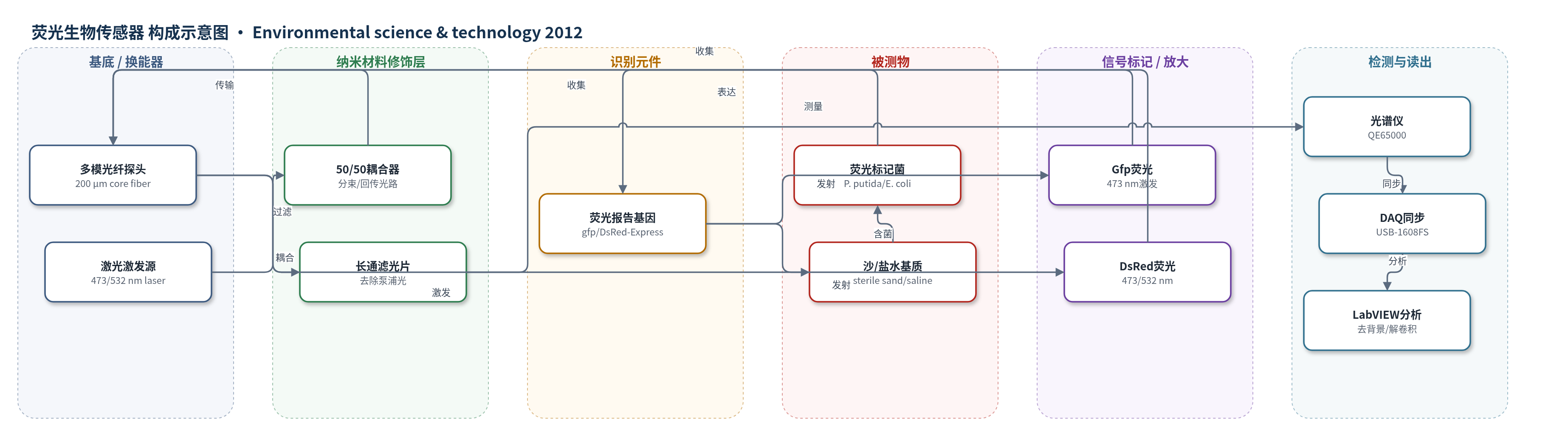
点击图片查看大图 · 依据论文自动绘制
传感器类型
荧光生物传感器
检测对象
荧光标记生物传感器菌(fluorescently labeled biosensor organisms,含 Gfp 标记 P. putida 852-137/E. coli-Gfp 与 DsRed 标记 E. coli-DsRed);样品基质:生理盐水(saline)与无菌沙(sterile sand)微宇宙。
检测原理
荧光标记生物传感器菌表达 Gfp 或 DsRed 报告蛋白,作为识别/报告元件。473 nm 或 532 nm 激光经多模光纤和 50/50 耦合器进入样品,激发荧光蛋白;Gfp 在 473 nm 激发下发射约 521/531 nm,DsRed 发射约 586 nm。同一光纤收集发射光,长通滤光片去除残余激发光,光谱仪测量积分强度。荧光强度与菌浓度或报告蛋白量成正比,稀释至 1/1024 仍保持线性(R^2=0.998)。混合样品用曲线拟合或 Alentsev-Fok 方法解卷积;时间分辨采集与 LabVIEW 实时扣除背景,并用指数衰减拟合校正光漂白。
检测灵敏度
LOD: 1.9 × 10^4 CFU/mL;E. coli-Gfp (saline): 5.7 × 10^4 CFU/mL;E. coli-Gfp (sand): 3.3 × 10^5 CFU/mL;E. coli-DsRed (saline, 473 nm): 3.0 × 10^6 CFU/mL;E. coli-DsRed (saline, 532 nm): 6.0 × 10^5 CFU/mL;P. putida 852−137 (saline): 4.8 × 10^5 CFU/mL;P. putida 852−137 (sand): 6.3 × 10^6 CFU/mL;线性范围: 1 × 10^9 CFU/mL 至 1/1024 稀释;R^2 = 0.998
效应效果
系统可在溶液和沙基质中识别、表征、区分并定量 Gfp 与 DsRed 标记菌,混合样品中通过曲线拟合和 Alentsev-Fok 方法解卷积,α 与稀释比在 log-log 图中呈线性。沙基质中可扣除背景自荧光,归一化光谱与溶液一致;积分强度较溶液降低约 3 倍,收集体积减少约 1/3,P. putida 852−137 在沙中检测限较盐水约降低 1 个数量级。高浓度样品 SNR 常大于 300,检测限按 SNR=3 外推。作者认为该方法适合生物修复中荧光标记降解菌的原位、非破坏性实时监测,并可扩展至荧光染料或标记化学物在土壤中的迁移监测。
传感器的构成
- 激发光源:100 mW 二极管泵浦固体激光器(473 nm、532 nm),用于激发 Gfp 和 DsRed 荧光蛋白。
- 光路控制:激光支架(OZ Optics)含电子快门,控制激发时序与持续时间。
- 传输/收集光纤:全硅 pyrocoat 高 OH- 多模非密封光纤(芯径 200 μm、包层 220 μm),传输激发光并收集荧光。
- 分束/耦合:50/50 耦合器,将激光分束导向样品并回传发射光。
- 滤光:长通滤光片,置于光谱仪前,去除残余泵浦光,仅保留荧光信号。
- 识别/报告元件:荧光蛋白报告基因(gfp、DsRed-Express)整合于生物传感器菌,响应细胞存在或基因表达并产生特征荧光。
- 换能/读出:光谱仪(QE65000, Ocean Optics)测量发射光谱;DAQ(USB-1608FS)同步快门与光谱仪;光电二极管(10530DAL)监测照明功率。
- 分析软件:LabVIEW 8.2 程序控制硬件,实时扣除背景并拟合或解卷积荧光光谱。
中文摘要
本研究提出一种新型时间分辨光纤 Optrode 系统,用于准确、实时、原位检测生物传感器生物体产生的荧光蛋白。该系统能够识别、表征、区分并定量红色和绿色荧光标记生物体,既适用于单独培养,也适用于混合水培养。在沙基质微宇宙中同样可实现检测,且信号强度的一致降低表明光纤收集体积约减少三分之一。Optrode 的灵敏度足以检测 1.9×10^4 CFU/mL 的荧光标记细胞浓度,适合生物修复试验中常见降解菌浓度范围。研究还表征了不同荧光蛋白的光漂白效应,发现其与细胞浓度呈线性关系,因此可在荧光采集与分析方法中予以校正。该工作为光纤原位荧光监测提供了概念验证,是建立完整、非破坏性原位监测系统以表征微生物活性和基因表达的重要步骤。
英文摘要
This research presents a novel, time-resolved fiber-optic "Optrode" system for accurate real-time in situ detection of fluorescent proteins produced by biosensor organisms. The Optrode fluorescence detection system was able to identify, characterize, differentiate, and quantify red and green fluorescently labeled organisms, individually and in mixed aqueous cultures. Detection was also possible in sand systems, where a consistent reduction in signal intensity indicates that signal collection volume was reduced by one-third. The optrode was shown to be sensitive enough to detect fluorescently labeled cell concentrations of 1.9 × 10(4) CFU/mL, indicating it is suitable for detecting typical concentrations of degrader organisms reported in bioremediation trials. The effect of fluorophore photobleaching was characterized for different fluorescent proteins and demonstrated a linear relationship to cell concentration, meaning the effect can be accounted for within methods of fluorescence collection and analysis. Proof of concept is provided for all aspects of this research, which represents an important step toward the goal of achieving a complete, nondestructive, in situ monitoring system to characterize all aspects of microbial activity and gene expression.