组成图示
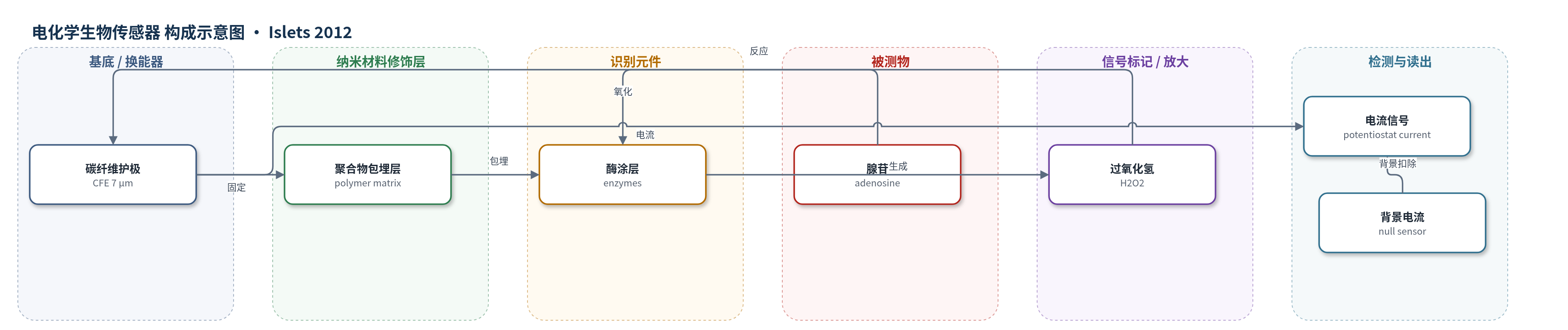
点击图片查看大图 · 依据论文自动绘制
传感器类型
电化学生物传感器
检测对象
腺苷(adenosine, Ado)、三磷酸腺苷(ATP);样品基质:孤立小鼠/大鼠胰腺胰岛周围灌注液(modified Krebs solution)
检测原理
腺苷从胰岛周围扩散进入碳纤维护极尖端的聚合物酶层,被固定酶催化转化为过氧化氢(H2O2)。H2O2 在碳纤维护极表面发生氧化反应,电子转移形成与腺苷浓度相关的电流。电位计记录电流,并与无酶空传感器背景差值结合,按标准曲线换算腺苷浓度。在 1–20 μM 范围内,电流随腺苷浓度近似线性增加。ATP 传感器采用类似酶促换能机制,检测 ATP 时需加入甘油作为共底物。该传感器依靠酶催化连续产生 H2O2 实现信号放大,微电极小体积和高界面响应使其适用于孤立胰岛的实时胞外腺苷监测。
检测灵敏度
线性范围: 1–20 mM(原文表述;正文图注为 μM)
效应效果
传感器在 1–20 μM 腺苷范围内准确,药物和营养物未直接干扰。小鼠 3 mM 葡萄糖基础腺苷 5.7 ± 0.6 μM(n=27,范围 1.2–11.6 μM);葡萄糖升至 5–25 mM 时腺苷下降,≥8 mM 后饱和,Hill K 约 4.3–4.6 mM、h=3。30 mM KCl 使腺苷升高约 3 倍至 16.4 ± 2.0 μM,依赖胞外 Ca2+;NTBI(50 μM)和 dipyridamole(10 μM)无显著影响。ATP 传感器未检出基础 ATP;外源 ATP 不改变小鼠腺苷,但升高大鼠腺苷,且被 POM-1 和 GMP 抑制。作者认为可用于小组织实时腺苷监测。
传感器的构成
- 基底/换能器电极:碳纤维护极(carbon fiber electrode, CFE),直径 7 μm(原文作 7 mm),传导电子并氧化 H2O2
- 修饰层:聚合物包埋层(polymer matrix),固定酶于电极尖端并允许腺苷扩散
- 识别/催化元件:酶涂层(enzymes),将腺苷催化转化为 H2O2
- 信号产物:过氧化氢(H2O2),在 CFE 表面氧化产生电流
- 对照元件:无酶空传感器(null sensor),不含酶,用于扣除背景电流
- 检测介质:改良 Krebs 溶液(modified Krebs solution),提供葡萄糖、KCl、Ca2+ 等灌注环境
- 辅助检测:ATP 酶传感器(ATP biosensor)与甘油(glycerol)共底物,用于 ATP 检测
中文摘要
朗格汉斯胰岛通过分泌胰岛素和胰高血糖素维持血糖稳态,其调节异常可导致糖尿病。腺苷是内源性嘌呤核苷,可激活 A1、A2A、A2B 和 A3 受体,外源腺苷能抑制胰岛素释放并促进胰高血糖素释放,但其在胰岛中的生理意义尚不清楚。本研究采用新型 7 μm 酶涂层碳纤维护极生物传感器,实时测量孤立小鼠和大鼠胰腺胰岛周围的腺苷水平。在 3 mM 葡萄糖条件下,小鼠胰岛基础腺苷水平约为 5.7 ± 0.6 μM;随着葡萄糖浓度升高,胞外腺苷下降,葡萄糖高于 8 mM 后抑制趋于饱和。30 mM KCl 使腺苷升高约 3 倍至 16.4 ± 2.0 μM,且该释放依赖胞外 Ca2+,提示腺苷经胞吐机制释放。此外,大鼠胰岛可将外源 ATP 转化为腺苷,而小鼠胰岛不能。该研究首次证明胰岛腺苷基础水平及其与胞外葡萄糖呈负相关。
英文摘要
The pancreatic islets of Langerhans are responsible for the regulated release of the endocrine hormones insulin and glucagon that participate in the control of glucose homeostasis. Abnormal regulation of these hormones can result in glucose intolerance and lead to the development of diabetes. Numerous efforts have been made to better understand the physiological regulators of insulin and glucagon secretion. One of these regulators is the purine nucleoside, adenosine. Though exogenous application of adenosine has been demonstrated to stimulate glucagon release and inhibit insulin release, the physiological significance of this pathway has been unclear. We used a novel 7 µm enzyme-coated electrode biosensor to measure adenosine levels in isolated rodent islets. In the mouse islets, basal adenosine levels in the presence of 3 mM glucose were estimated to be 5.7 ± 0.6 µM. As glucose was increased, extracellular adenosine diminished. A 10-fold increase of extracellular KCl increased adenosine levels to 16.4 ± 2.0 µM. This release required extracellular Ca (2+) suggesting that it occurred via an exocytosis-dependent mechanism. We also found that while rat islets were able to convert exogenous ATP into adenosine, mouse islets were unable to do this. Our study demonstrates for the first time the basal levels of adenosine and its inverse relationship to extracellular glucose in pancreatic islets.