组成图示
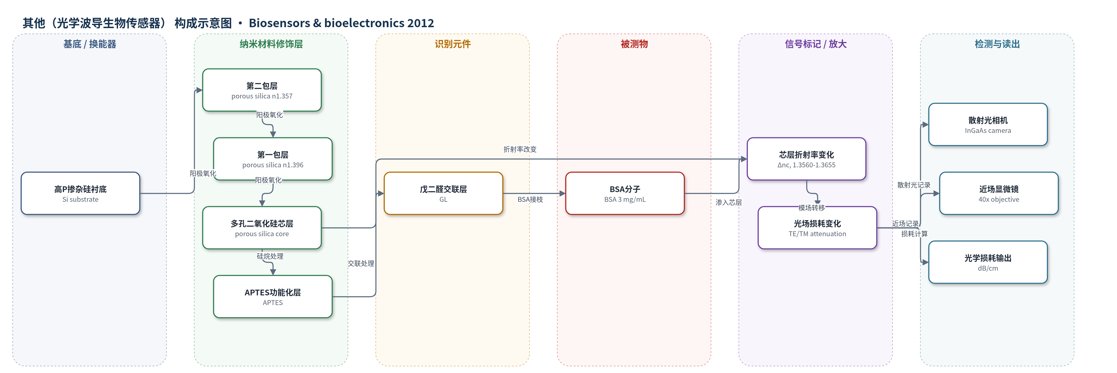
点击图片查看大图 · 依据论文自动绘制
传感器类型
其他(光学波导生物传感器)
检测对象
牛血清白蛋白(BSA, bovine serum albumin),样品基质:BSA 水溶液(3 mg/mL)
检测原理
该传感器采用多孔二氧化硅平面 ARROW 波导作为换能器。1.55 μm 激光以 TE 或 TM 偏振耦合进入多孔芯层,光场由芯层上表面全反射和芯层/第一包层界面的反谐振反射共同限制。APTES 和戊二醛功能化后,BSA 分子渗入高孔隙率芯层,使芯层有效折射率由 nc0 增加 Δnc。当 Δnc≥0 时,芯层/第一包层界面反射率 R 显著升高,传播模从第一包层转向芯层,导致波导光学损耗降低。实验以表面散射光强度沿传播方向的指数衰减斜率计算损耗,并用近场显微镜观察模场分布。多孔结构提供高比表面,使光场与渗入分子重叠增强,属于无标记光学折射率传感,无酶或核酸放大。
检测灵敏度
原文未报告 LOD、线性范围、灵敏度斜率或相关系数。
效应效果
实验验证了氧化多孔硅 ARROW 作为无标记光学生物换能器的可行性。BSA 接枝使 TE 和 TM 偏振的光学衰减分别由约 5 dB/cm 和 7 dB/cm 降至约 3 dB/cm 和 3.7 dB/cm,降低约 2 dB/cm 和 3 dB/cm;相对氧化后约 7.5 dB/cm 和 10 dB/cm 也明显下降。近场模场在 BSA 接枝后几乎完全限制在芯层内,与 FD-BPM 模拟一致。作者指出多孔硅 ARROW 相比传统倏逝波结构可提高光场与分子重叠,适合无标记生物分子检测;但本文未报告选择性、抗干扰、RSD、实际样品回收率或与 ELISA/HPLC/qPCR 的对比,后续拟集成 Mach-Zehnder 干涉仪并测试不同浓度分析物。
传感器的构成
- 基底:高 P 掺杂硅衬底(Si substrate, 7 mΩ cm),提供机械支撑与阳极氧化基底。
- 第二包层:多孔二氧化硅第二包层(porous silica second cladding, 2 μm, 21% porosity, n≈1.357),参与反谐振反射。
- 第一包层:多孔二氧化硅第一包层(porous silica first cladding, 1.4 μm, 11% porosity, n≈1.396),高折射率低孔层,增强反谐振反射。
- 多孔芯层:多孔二氧化硅芯层(porous silica core, 3.8 μm, 32% porosity, n≈1.298),高孔隙率传感区,容纳 BSA 并改变有效折射率。
- 硅烷功能化层:3-氨基丙基三乙氧基硅烷(APTES)处理层,在多孔表面引入氨基。
- 交联固定层:戊二醛(GL, glutaraldehyde)处理层,交联固定 BSA。
- 生物分子接枝层:牛血清白蛋白(BSA, 3 mg/mL)渗入多孔芯层,作为模型生物分子产生折射率变化。
中文摘要
本文报道了一种基于多孔硅(PS)反谐振反射光学波导(ARROW)的光学生物传感器换能器。与传统倏逝波生物传感器相比,ARROW 结构通过使传播光场与渗入多孔芯层的分子更好地重叠,从而提高分子相互作用灵敏度。本研究旨在考察采用 ARROW 结构的光学生物传感器工作模式,并在给定折射率范围内,于待测分子接枝前对反谐振条件进行调节。所研究 ARROW 结构由同一多孔硅材料经单步电化学阳极氧化制备;经氧化和化学功能化处理后,牛血清白蛋白(BSA)主要接枝在上层。模拟结果表明,该传感器在折射率 1.3560-1.3655 范围内工作。实验上通过传播光场和损耗变化实现生物分子的光学检测。结果显示,生物分子接枝后光学衰减降低,对应芯层折射率变化 Δnc;TE 和 TM 偏振下衰减分别降低约 2 dB/cm 和 3 dB/cm。检测阶段光场几乎位于芯层内,与模拟近场分布一致。
英文摘要
Recently, we demonstrated that Anti Resonant Reflecting Optical Waveguide (ARROW) based on porous silicon (PS) material can be used as a transducer for the development of a new optical biosensor. Compared to a conventional biosensor waveguide based on evanescent waves, the ARROW structure is designed to allow a better overlap between the propagated optical field and the molecules infiltrated in the porous core layer and so to provide better molecular interactions sensitivity. The aim of this work is to investigate the operating mode of an optical biosensor using the ARROW structure. We reported here an extensive study where the antiresonance conditions were adjusted just before the grafting of the studied molecules for a given refractive index range. The interesting feature of the studied ARROW structure is that it is elaborated from the same material which is the porous silicon obtained via a single electrochemical anodization process. After oxidation and preparation of the inner surface of porous silicon by a chemical functionalization process, bovine serum albumin (BSA) molecules, were attached essentially in the upper layer. Simulation study indicates that the proposed sensor works at the refractive index values ranging from 1.3560 to 1.3655. The experimental optical detection of the biomolecules was obtained through the modification of the propagated optical field and losses. The results indicated that the optical attenuation decreases after biomolecules attachment, corresponding to a refractive index change Δn(c) of the core. This reduction was of about 2 dB/cm and 3 dB/cm for Transverse Electric (TE) and Transverse Magnetic (TM) polarizations respectively. Moreover, at the detection step, the optical field was almost located inside the core layer. This result was in good agreement with the simulated near field profiles.