组成图示
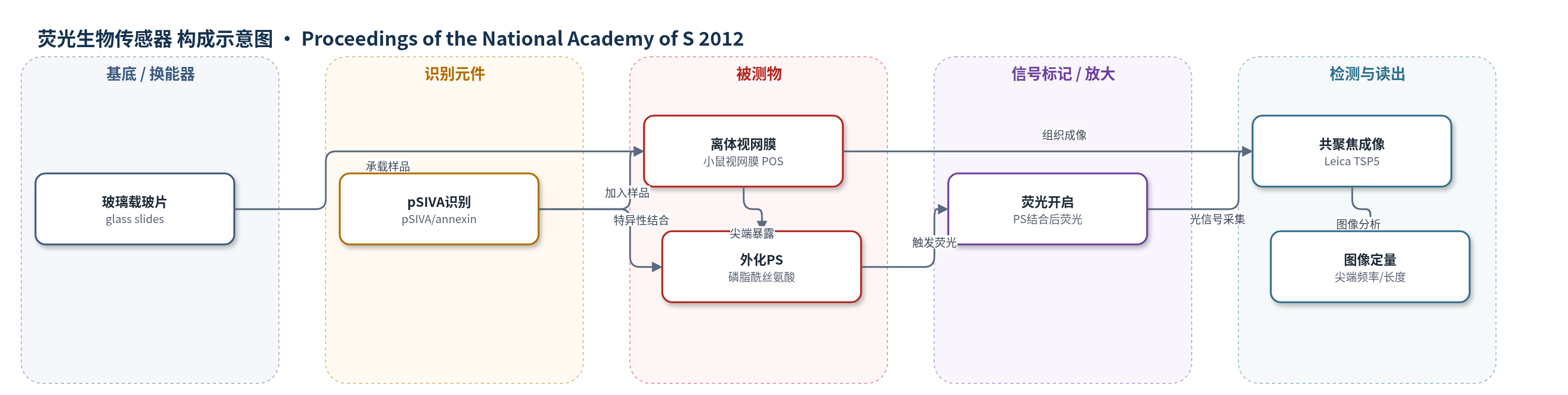
点击图片查看大图 · 依据论文自动绘制
传感器类型
荧光生物传感器
检测对象
磷脂酰丝氨酸(phosphatidylserine, PS);样品基质为离体小鼠视网膜、分离光感受器外段(POS)颗粒及 RPE 培养体系
检测原理
pSIVA 是一种 annexin 基荧光开关生物传感器,其识别元件可特异性结合细胞膜外叶暴露的磷脂酰丝氨酸(PS)。在正常膜中 PS 主要位于胞质叶,当 POS 尖端发生 PS 外化时,pSIVA 与外化 PS 结合,引起其荧光状态由关闭或低荧光转变为开启或高荧光。由于 pSIVA 无需洗脱或固定即可用于活体组织,PS 暴露面积越大、暴露尖端越多,结合 pSIVA 产生的荧光信号越强,荧光标记的 POS 尖端频率和长度也越高。激光共聚焦显微镜采集 pSIVA 荧光图像,通过图像分析定量 PS 标记尖端,从而反映 POS 尖端 PS 暴露水平及其昼夜变化。
检测灵敏度
原文未报告 LOD、线性范围、灵敏度斜率或相关系数。
效应效果
pSIVA 对 PS 高度特异,可在活体离体小鼠视网膜中无需洗脱和固定即清晰界定 POS 尖端,结果与 FITC-A5、Alexa Fluor 488-A5 和 αPS 一致。竞争实验显示 αPS 和 A5 竞争结合 POS,而视紫红质抗体和 β-半乳糖苷酶无竞争。PS 阻断使 RPE-J 细胞对 POS 颗粒的结合分别降低 46% 和 45%,内化也降低;磷脂酶 C 预处理增强 PS 识别后 RPE 结合和吞噬增强,非磷脂酶处理则降低。野生型小鼠光启动后 PS 标记尖端频率和长度达峰,尖端长度较光启动前后 1 h 平均增加 43%;Itgb5−/− 和 Mfge8−/− 小鼠无昼夜变化,P<0.05。作者认为该方法适用于实时监测活体视网膜 PS 外化及吞噬节律。
传感器的构成
- 样品承载基底:玻璃载玻片(glass slides),承载离体视网膜或 POS 样品
- 识别元件:pSIVA(polarity-sensitive indicator of viability and apoptosis),annexin 基生物传感器,特异性识别外化磷脂酰丝氨酸(PS)
- 信号标记物:pSIVA 内禀荧光开关,结合 PS 后荧光状态改变,提供可成像信号
中文摘要
在哺乳动物视网膜中,光感受器外段(POS)的终身更新依赖昼夜节律性远端杆状 POS 尖端脱落,以及随后视网膜色素上皮(RPE)在光启动后对其吞噬。促进或同步 POS 尖端脱落的分子机制尚不清楚。本研究通过抗体、annexin V 和 pSIVA(polarity-sensitive indicator of viability and apoptosis,一种具有可切换荧光状态的 annexin 基生物传感器)定量活体 POS 的膜不对称性,检测磷脂酰丝氨酸(PS)的表面暴露。结果显示,分离的 POS 颗粒具有外化 PS,阻断或去除 PS 会降低其在培养中与 RPE 的结合和吞噬。对新鲜离体小鼠视网膜活体成像发现,PS 外化局限于 POS 尖端且边界清晰。野生型小鼠中,暴露 PS 的杆尖端频率及 PS 暴露尖端长度在光启动后不久达到峰值;而在缺乏 RPE 昼夜吞噬节律的 Itgb5−/− 或 Mfge8−/− 小鼠中,PS 标记的 POS 尖端不随时间变化。这些数据表明,局部 PS 暴露是杆状 POS 尖端表面的特异性分子标志,PS 暴露增强先于脱落和吞噬,提示表面 PS 促进这些过程,并且 PS 标记 POS 尖端的昼夜节律需要 RPE 活性参与。
英文摘要
In the mammalian retina, life-long renewal of light-sensitive photoreceptor outer segments (POS) involves circadian shedding of distal rod POS tips and their subsequent phagocytosis by the adjacent retinal pigment epithelium (RPE) every morning after light onset. Molecular mechanisms that promote or synchronize POS tip shedding have thus far remained unknown. Here we examined plasma membrane asymmetry of living POS by quantifying surface exposure of the membrane phospholipid phosphatidylserine (PS) using antibodies, annexin V, and pSIVA (polarity-sensitive indicator of viability and apoptosis), an annexin-based biosensor with switchable states of fluorescence. We found that isolated POS particles possess externalized PS, whose blockade or removal reduces their binding and engulfment by RPE in culture. Imaging of live photoreceptors in freshly dissected mouse retina detected PS externalization restricted to POS tips with discrete boundaries. In wild-type mice, frequency of rod tips exposing PS and length of tips with exposed PS peak shortly after light onset. In contrast, PS-marked POS tips do not vary in mice lacking the diurnal phagocytic rhythm of the RPE due to loss of either the phagocytosis receptor αvβ5 integrin, expressed by the RPE but not by photoreceptors, or its extracellular ligand milk fat globule-EGF factor 8 (MFG-E8). These data identify a molecular distinction, localized PS exposure, that is specific to the surface of rod POS tips. Enhanced PS exposure preceding rod shedding and phagocytosis suggests that surface PS promotes these processes. Moreover, our results demonstrate that the diurnal rhythm of PS demarcation of POS tips is not intrinsic to rod photoreceptors but requires activities of the RPE as well.