组成图示
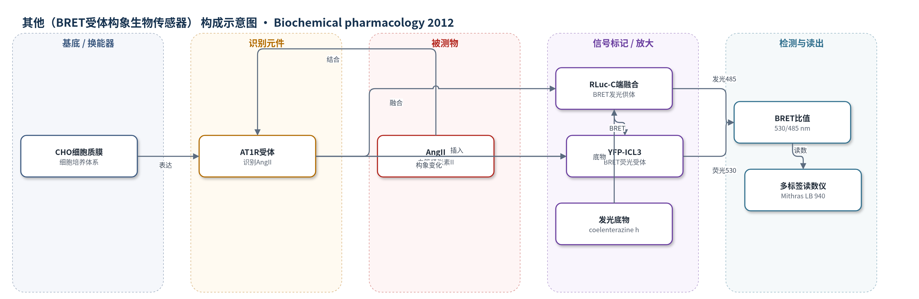
点击图片查看大图 · 依据论文自动绘制
传感器类型
其他(BRET受体构象生物传感器)
检测对象
血管紧张素II(angiotensin II, AngII);样品基质:CHO细胞培养体系/细胞膜
检测原理
AngII与AT1R结合后,受体发生构象变化。AT1R-BS中YFP插入第三胞内环、RLuc融合于C端,二者构成BRET供体-受体对。RLuc在coelenterazine h存在下发光,能量非辐射转移至YFP,YFP在530 nm发射荧光;BRET比值定义为530 nm荧光/485 nm发光。当AngII直接激活AT1R-BS时,受体构象改变使YFP与RLuc距离或取向变化,BRET信号下降;在AT1R同源二聚体中,一个亚基被AngII激活可通过DRY基序介导的变构通讯改变另一亚基构象,导致早期BRET上升并促进β-arrestin2结合。DRY/AAY突变破坏激活亚基完全活性构象,消除上述变构信号。该传感器无酶促放大,依赖分子内BRET效率变化反映受体激活状态。
检测灵敏度
原文未报告LOD、线性范围、灵敏度斜率或相关系数。
效应效果
BRET滴定显示AT1R-RLuc/AT1R-YFP呈饱和曲线,PM-YFP呈线性,证明二聚化特异性。10 μM candesartan阻断敏感亚基后,100 nM AngII选择性刺激CR-AT1R;2 μM U-73122不阻断非刺激亚基β-arrestin2结合,提示不依赖PLC。DRY/AAY突变消除β-arrestin2结合、构象变化和负协同。4°C下0.01 nM 125I-AngII与1 mM冷AngII解离实验显示WT-AT1R负协同,DRY/AAY-AT1R无此效应。数据为3–8次实验均值±SEM,p<0.001。作者认为该结果揭示AT1R二聚体变构机制及DRY基序关键作用,为药理学干预提供依据。
传感器的构成
- 基底/细胞体系:CHO细胞质膜,承载AT1R并维持受体激活与BRET测量环境
- 识别元件:1型血管紧张素受体(AT1R),结合AngII并发生构象变化;CR-AT1R、DRY/AAY-AT1R用于选择性刺激与机制研究
- 发光供体标记:海肾荧光素酶(RLuc)融合于AT1R C端,在coelenterazine h存在下发光,作为BRET供体
- 荧光受体标记:黄色荧光蛋白(YFP)插入AT1R第三胞内环(N231-K232之间),作为BRET受体,其荧光随受体构象改变而变化
- 信号底物:coelenterazine h,RLuc发光底物,激发BRET信号
- 读出装置:Mithras LB 940多标签读数仪,测量485 nm发光与530 nm荧光,计算BRET ratio
中文摘要
G蛋白偶联受体(GPCR)二聚化显著影响受体信号多样性,二聚体亚基间的变构通讯可改变配体结合、受体构象及与不同效应蛋白的相互作用。本研究在CHO细胞中瞬时表达野生型和突变型1型血管紧张素受体(AT1R)同源二聚体,研究其亚基间变构相互作用。实验通过选择性刺激一个亚基,监测另一亚基的β-arrestin2结合和构象变化。β-arrestin2与非刺激亚基的相互作用采用生物发光共振能量转移(BRET)方法监测;为直接测量非刺激亚基构象变化,作者构建了BRET分子内受体生物传感器,将黄色荧光蛋白(YFP)插入AT1R第三胞内环,并在C端融合海肾荧光素酶(RLuc)。结果检测到非刺激亚基的β-arrestin2结合和构象改变,放射性配体解离实验还观察到同源二聚体配体结合的负协同性。突变激活亚基中保守DRY序列后,所有变构效应消失。这些结果表明AT1R同源二聚体中的变构相互作用显著影响非刺激亚基功能,DRY基序在其中起关键作用。
英文摘要
G protein coupled receptor (GPCR) dimerization has a remarkable impact on the diversity of receptor signaling. Allosteric communication between the protomers of the dimer can alter ligand binding, receptor conformation and interactions with different effector proteins. In this study we investigated the allosteric interactions between wild type and mutant protomers of type 1 angiotensin receptor (AT₁R) dimers transiently expressed in CHO cells. In our experimental setup, one protomer of the dimer was selectively stimulated and the β-arrestin2 binding and conformation alteration of the other protomer was followed. The interaction between β-arrestin2 and the non-stimulated protomer was monitored through a bioluminescence resonance energy transfer (BRET) based method. To measure the conformational alterations in the non-stimulated protomer directly, we also used a BRET based intramolecular receptor biosensor, which was created by inserting yellow fluorescent protein (YFP) into the 3rd intracellular loop of AT₁R and fusing Renilla luciferase (RLuc) to its C terminal region. We have detected β-arrestin2 binding, and altered conformation of the non-stimulated protomer. The cooperative ligand binding of the receptor homodimer was also observed by radioligand dissociation experiments. Mutation of the conserved DRY sequence in the activated protomer, which is also required for G protein activation, abolished all the observed allosteric effects. These data suggest that allosteric interactions in the homodimers of AT₁R significantly affect the function of the non-stimulated protomer, and the conserved DRY motif has a crucial role in these interactions.