组成图示
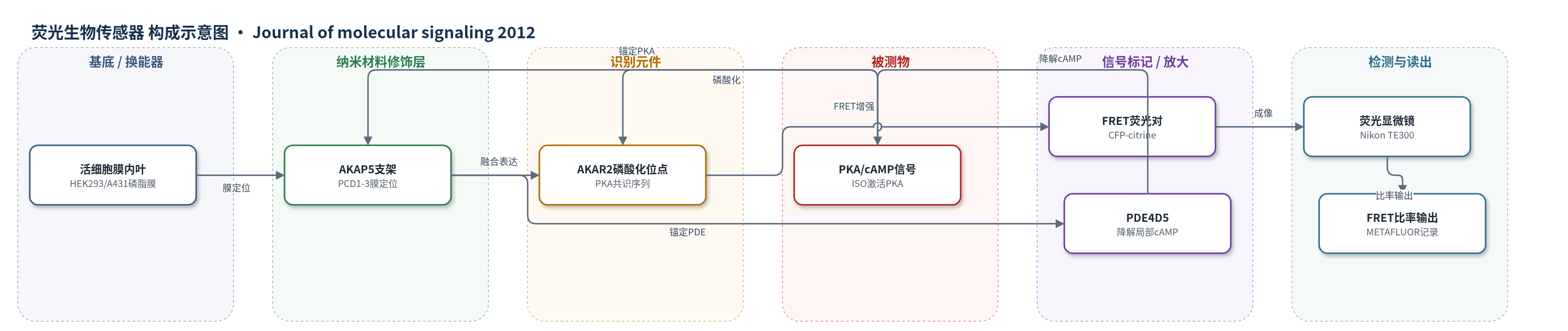
点击图片查看大图 · 依据论文自动绘制
传感器类型
荧光生物传感器
检测对象
蛋白激酶A活性/磷酸化事件(PKA activity / PKA-catalyzed phosphorylation)、局部环磷酸腺苷(cAMP);样品基质:HEK293/A431活细胞,β-肾上腺素激动剂ISO刺激
检测原理
β-肾上腺素激动剂ISO激活膜上β2-肾上腺素受体和腺苷酸环化酶,使细胞内cAMP升高并激活PKA。AKAP5通过PCD1-3定位于膜内叶,其RII结合域将PKA锚定在受体附近,使PKA优先磷酸化融合在N端的AKAR2共识序列。AKAR2磷酸化后,Forkhead同源域的磷酸氨基酸结合序列发生构象变化,拉近CFP与citrine,使YFP/CFP FRET比率升高。该比率随局部cAMP/PKA活性增强而升高。AKAP5同时锚定PDE4D5,后者降解局部cAMP,降低PKA活性并促使AKAR2去磷酸化,使FRET信号在约60秒内回落。因此传感器通过支架局部富集、PKA磷酸化识别和FRET换能实现实时空间分辨读出,PDE4D5提供负反馈以塑造信号动力学。
检测灵敏度
原文未报告 LOD、线性范围、灵敏度斜率或 R^2。
效应效果
AKAR2-AKAP5在HEK293中保留天然AKAP5功能:AKAP5敲低抑制ISO诱导ERK1/2激活,表达融合蛋白可完全恢复。FRET信号在HEK293约10秒达峰、60秒回基线;A431中延迟且幅度低。AKAR2-AKAP12呈渐进升高且无明显逆转。PKA抑制剂H89(10 μM)、KT5720(1 μM)及Ht-31肽(50 μM)消除FRET,对照Ht-31p不消除。PDE4D5敲低、Rolipram(10 μM)或Δ1-145,T/P395不改变初始峰,但消除快速逆转。实验重复至少三次,p<0.05。作者认为该传感器可实时解析AKAP支架对PKA/cAMP信号的时空塑造。
传感器的构成
- 细胞基底:HEK293或A431活细胞,提供膜内叶磷脂环境和胞内cAMP/PKA信号微环境
- 支架定位层:AKAP5(AKAP79/75)的PCD1-3正电荷结构域,静电结合膜内叶磷脂,将传感器锚定于膜附近
- 支架识别层:AKAP5的RII结合域与PDE4D结合域,分别锚定PKA和PDE4D5,形成局部信号支架
- 磷酸化识别元件:AKAR2模块,含Forkhead同源域磷酸氨基酸结合序列和PKA共识磷酸化位点
- 荧光换能器:CFP供体与citrine(YFP变体)受体,磷酸化改变二者距离,产生FRET比率
- 信号调控元件:PDE4D5,降解局部cAMP,加速PKA去磷酸化和信号逆转
- 读出系统:Nikon Eclipse TE300显微镜、ORCA-ER CCD相机和METAFLUOR 7.2软件,记录YFP/CFP比率
中文摘要
蛋白激酶A(PKA)是细胞信号转导的关键调控酶,可磷酸化多种靶蛋白,并被A-激酶锚定蛋白(AKAP)家族在空间上锚定。AKAR2是一种活细胞FRET生物传感器,当被PKA磷酸化时产生荧光共振能量转移信号。AKAP5是AKAP家族重要成员,可锚定PKA、磷酸二酯酶PDE4D及G蛋白偶联受体(GPCR),并介导GPCR向ERK1/2的MAPK级联激活。本研究构建AKAR2-AKAP5融合生物传感器,实时监测β-肾上腺素刺激下AKAP5被PKA磷酸化的时空动态。结果显示,AKAR2-AKAP5报告的PKA激活比AKAR2-AKAP12更快且空间更局限,表明AKAP5对局部PKA激活具有时空限制并塑造信号响应。锚定于AKAP5的PDE4D可在约60秒内逆转β-肾上腺素激动剂引起的细胞内cAMP升高;而AKAP12不能在该时间窗内衰减cAMP升高。将AKAP5的PDE4D结合域融合至AKAP12可加速cAMP积累的逆转。结论表明,携带锚定酶的AKAP支架可塑造细胞信号的时间与空间特征。
英文摘要
BACKGROUND: PKA, a key regulator of cell signaling, phosphorylates a diverse and important array of target molecules and is spatially docked to members of the A-kinase Anchoring Protein (AKAP) family. AKAR2 is a biosensor which yields a FRET signal in vivo, when phosphorylated by PKA. AKAP5, a prominent member of the AKAP family, docks several signaling molecules including PKA, PDE4D, as well as GPCRs, and is obligate for the propagation of the activation of the mitogen-activated protein kinase cascade from GPCRs to ERK1,2.
RESULTS: Using an AKAR2-AKAP5 fusion "biosensor", we investigated the spatial-temporal activation of AKAP5 undergoing phosphorylation by PKA in response to β-adrenergic stimulation. The pattern of PKA activation reported by AKAR2-AKAP5 is a more rapid and spatially distinct from those "sensed" by AKAR2-AKAP12. Spatial-temporal restriction of activated PKA by AKAP5 was found to "shape" the signaling response. Phosphatase PDE4D tethered to AKAP5 also later reverses within 60 s elevated intracellular cyclic AMP levels stimulated by β-adrenergic agonist. AKAP12, however, fails to attenuate the rise in cyclic AMP over this time. Fusion of the AKAP5 PDE4D-binding-domain to AKAP12 was found to accelerate a reversal of accumulation of intracellular cyclic AMP.
CONCLUSION: AKAPs, which are scaffolds with tethered enzymes, can "shape" the temporal and spatial aspects of cell signaling.