组成图示
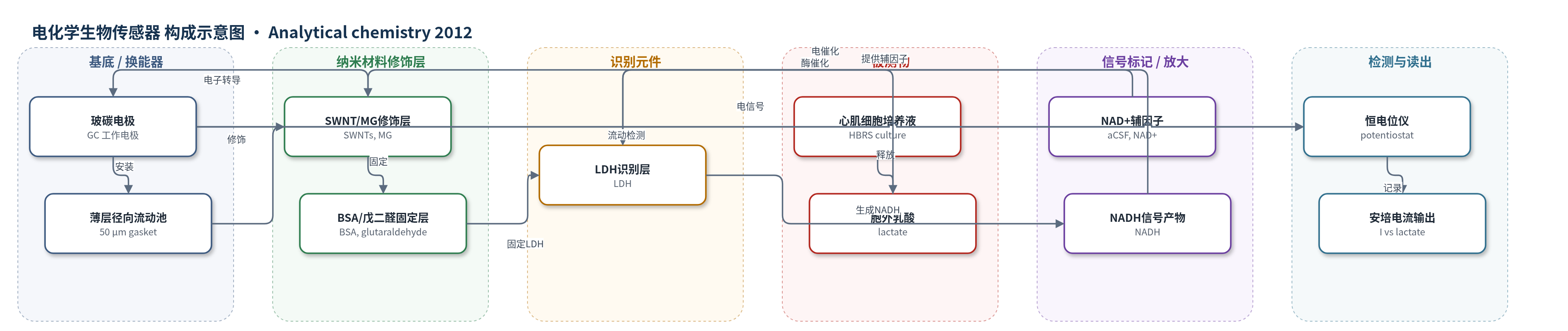
点击图片查看大图 · 依据论文自动绘制
传感器类型
电化学生物传感器
检测对象
乳酸(lactate);样品基质:新生大鼠心肌细胞胞外培养液(HBRS 细胞培养液)
检测原理
心肌细胞缺氧时糖酵解增强,胞外乳酸增加。FEP 管以负压从培养瓶抽取心肌细胞培养液,与含 2.0 mM NAD+ 的 aCSF 在 T 型接头在线混合后进入薄层径向电化学流动池。LDH 识别层催化乳酸与 NAD+ 反应生成丙酮酸和 NADH;NADH 在 MG/SWNT 修饰的 GC 电极上于 0.0 V 被电催化氧化,电子经 SWNTs 转导至 GC,产生与乳酸浓度成正比的安培电流。外部 NAD+ 灌注既补充辅因子,又缓冲缺氧引起的 pH 变化;MG/SWNT 低电位电催化降低内源性电活性物质干扰。
检测灵敏度
LOD: 0.16 mM (S/N = 3);线性范围: 0.20–10 mM;I (nA) = 25.6 C_Lactate (mM) + 20.1,γ = 0.996
效应效果
该方法对心肌细胞培养液中的内源性电活性物质具有良好选择性:10 µM 尿酸(UA)、10 µM 肾上腺素(E)、10 µM 去甲肾上腺素(NE)和 5 µM 抗坏血酸(AA)均无可记录电流响应;向培养液中加入乳酸氧化酶(LOx)消耗乳酸后电流回落至基线,表明对 H2O2 及缺氧产生的 ROS、黄嘌呤、次黄嘌呤、丙二醛等无干扰,且耐受缺氧引起的 pH 变化。系统连续监测 2.0 mM 乳酸至少 2 h 稳定,间断使用 8 天仍保持响应,重复测量 RSD 为 1.53%(n=5)。在细胞密度约 0.5×10^3 cells/mm2 时,生理胞外乳酸为 1.1±0.1 mM(n=3);15 µM FCCP 诱导缺氧 20 min 后升至 255±30.3%(n=3)。作者认为该平台可用于心肌生理与病理能量代谢研究。
传感器的构成
- 基底/换能器电极:玻碳电极(GC, glassy carbon electrode)作为工作电极,不锈钢辅助电极和 Ag/AgCl 参比电极构成薄层径向电化学流动池(thin-layer radial electrochemical flow cell)
- 纳米材料修饰层:单壁碳纳米管(SWNTs)负载亚甲蓝(MG, methylene green),作为 NADH 电催化氧化的电子转导/电催化层
- 识别元件:乳酸脱氢酶(LDH, lactate dehydrogenase)催化乳酸氧化为丙酮酸,并将 NAD+ 还原为 NADH
- 固定/封闭层:牛血清白蛋白(BSA)与戊二醛(glutaraldehyde)用于将 LDH 固定于 MG/SWNT 修饰电极表面
- 辅因子灌注层:人工脑脊液(aCSF)中 2.0 mM NAD+ 外部灌注,在线混合培养液,补充 LDH 辅因子并缓冲 pH
- 采样/流动层:FEP 管负压驱动采样心肌细胞培养液,经 T 型接头与 NAD+ 溶液混合后进入流动池
- 信号产物:NADH 由 LDH 催化生成,在 MG/SWNT 修饰 GC 电极上于 0.0 V 被电催化氧化,产生安培电流
- 读出装置:恒电位仪与计算机(potentiostat & computer)记录电流信号
中文摘要
连续监测心肌细胞乳酸生成对心肌能量代谢及心肌缺氧/缺血诊断治疗具有重要意义。本研究报道了一种基于脱氢酶的电化学生物传感器与负压驱动培养液采样联用,用于连续监测心肌缺氧后新生大鼠心肌细胞胞外乳酸生成。为消除缺氧引起 pH 变化对传感器响应的影响,并补充乳酸脱氢酶(LDH)反应所需烟酰胺腺嘌呤二核苷酸(NAD+)辅因子,将含 NAD+ 的人工脑脊液(aCSF)外部灌注,并在检测前与细胞培养液在线混合。该方法对心肌细胞胞外培养液中内源性电活性物质具有高选择性,并对缺氧后 pH 变化具有良好耐受性。乳酸动态线性范围为 0.20–10 mM(I(nA)=25.6 C_Lactate(mM)+20.1,γ=0.996),检出限 0.16 mM(S/N=3)。生理状态下胞外乳酸为 1.1±0.1 mM(n=3,细胞密度约 0.5×10^3 cells/mm2);FCCP 诱导缺氧 20 min 后升至生理水平的 255±30.3%(n=3)。该平台可用于研究心脏生理和病理过程中的能量代谢。
英文摘要
Continuous monitoring of lactate production from cardiomyocytes is of great physiological and pathological importance since the level of lactate in extracellular fluid is closely associated with myocardial energy metabolism with implication in the diagnosis and therapeutics of myocardial hypoxia and ischemia. This study demonstrates an electrochemical approach to continuous monitoring of lactate production from neonatal rat cardiomyocytes following myocardial hypoxia with a dehydrogenase-based electrochemical biosensor and a negative pressure driven culture sampling. To eliminate the effect of pH variation occurring following the cardiomyocyte hypoxia on the biosensor response and to supply nicotinamide adenine dinucleotide (NAD(+)) cofactor necessary for the enzymatic reaction of lactate dehydrogenase (LDH), artificial cerebrospinal fluid (aCSF) containing NAD(+) cofactor is externally perfused and mixed online with cell culture before the culture goes to the detector. The method exhibits a high selectivity against the electrochemically active species endogenously existing in the extracellular culture of cardiomyocytes and a high tolerance against the variation of pH following cardiomyocyte hypoxia. The dynamic linear range for lactate detection is from 0.20 to 10 mM (I (nA) = 25.6 C(Lactate) (mM) + 20.1, γ = 0.996) with a detection limit of 0.16 mM (S/N = 3). The physiological level of the extracellular lactate of neonatal rat cardiomyocytes is determined to be 1.1 ± 0.1 mM (n = 3) with the cell density of about 0.5 × 10(3) cells/mm(2). When the cardiomyocytes are subject to hypoxia induced with anoxic reagents, carbonyl cyanide 4-(trifluoromethoxy)phenylhydrazone (FCCP), the extracellular lactate increases to 255 ± 30.3% (n = 3), relative to the physiological level, following 20 min of the hypoxia. This study essentially offers a new and effective electrochemical platform for investigating energy metabolism during cardiac physiological and pathological processes.