组成图示
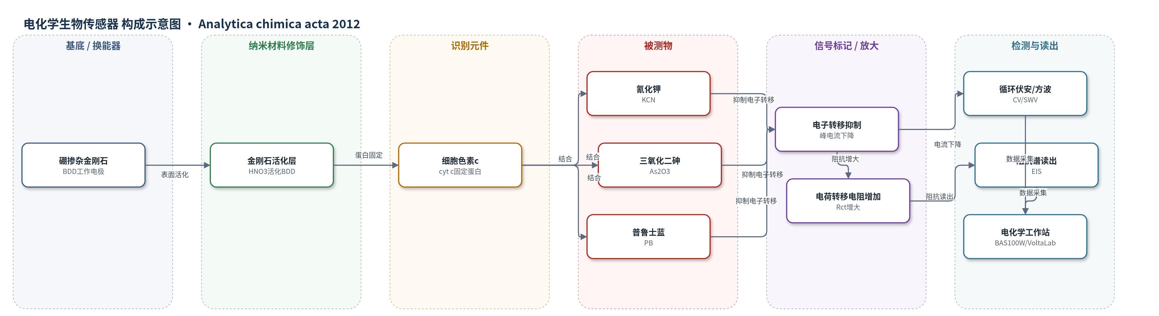
点击图片查看大图 · 依据论文自动绘制
传感器类型
电化学生物传感器
检测对象
普鲁士蓝(Prussian blue, PB, Fe2K(CN)12·16H2O)、氰化钾(potassium cyanide, KCN)、三氧化二砷(arsenic trioxide, As2O3);样品基质为0.1 M磷酸盐缓冲液(PBS,pH 7.0)
检测原理
该传感器以硼掺杂金刚石(BDD)电极为换能器,表面固定细胞色素c(cyt c)。cyt c保留天然电子传递能力,在0.1 M PBS(pH 7)中于约−240 mV出现不可逆还原峰。氰化钾(KCN)、三氧化二砷(As2O3)和普鲁士蓝(PB)通过静电/配位作用结合到cyt c表面正电氨基酸侧链,而非血红素铁位点,阻碍蛋白与BDD之间的电子转移。被测物浓度越高,结合量越大,循环伏安(CV)和方波伏安(SWV)还原峰电流越低;电化学阻抗谱(EIS)中界面电荷转移电阻Rct和界面电容增大,可用Randles等效电路定量。该体系无酶催化或核酸放大,依赖蛋白直接电子转移和BDD低背景、宽电位窗实现检测。
检测灵敏度
LOD: 4.3–9.1 μM(摘要);灵敏度: (1.1–4.5) × 10−8 A μM−1(摘要);CV(KCN) LOD: 4.22 μM,线性范围: 0–2 μM,灵敏度: 3.9 × 10⁻⁸ A μM−1;CV(As) LOD: 8.08 μM,线性范围: 0–4 μM,灵敏度: 2.1 × 10⁻⁸ A μM−1;CV(PB) LOD: 9.09 μM,线性范围: 0–4 μM,灵敏度: 1.8 × 10⁻⁸ A μM−1;SWV(KCN) LOD: 9.08 μM,线性范围: 0–10 μM,灵敏度: 4.5 × 10⁻⁸ A μM−1;SWV(As) LOD: 22.02 μM,线性范围: 0–10 μM,灵敏度: 1.9 × 10⁻⁸ A μM−1;SWV(PB) LOD: 37.49 μM,线性范围: 0–6 μM,灵敏度: 1.1 × 10⁻⁸ A μM−1;EIS LOD: KCN 9.97 μM、As 8.99 μM、PB 9.00 μM,EIS 灵敏度: 0.5(原文表头单位显示为 μ μM−1);UV–vis 相关系数: 0.99(结合曲线)
效应效果
该传感器在PBS中对KCN、As2O3和PB呈结合依赖响应,CV与SWV精度经F检验等价(PB标准差分别为1.36×10⁻⁸和1.19×10⁻⁸,F=0.773,低于Fcrit=5.05)。EIS表明固定cyt c后BDD电催化优势未丧失,Rct和界面电容随浓度增加。UV–vis摩尔吸光系数为KCN 4.05 cm mM−1、PB 2.43 cm mM−1、As2O3 1.59 cm mM−1,显示对KCN亲和最高。作者称检出限低于EPA/WHO指南(氰化物200 μg/L、砷10 μg/L),结论给出氰化物0.27–55 mg/L、砷1.58–4.31 mg/L。未报告实际样品回收率、长期稳定性或RSD。
传感器的构成
- 基底/换能器电极:硼掺杂金刚石(BDD)电极,经Al2O3抛光和1 M HNO3活化,作为工作电极提供低背景、宽电位窗电子转移动态界面
- 识别元件:细胞色素c(cytochrome c, cyt c,horse heart Type I),经孵育固定于BDD表面,作为生物识别蛋白和电子传递中心
- 被测物:氰化钾(potassium cyanide, KCN)、三氧化二砷(arsenic trioxide, As2O3)、普鲁士蓝(Prussian blue, PB, Fe2K(CN)12·16H2O),与cyt c表面正电侧链结合
- 参比/对电极:Ag/AgCl(3 M)参比电极和铂丝对电极,构成三电极电化学测量体系
- 缓冲介质:0.1 M磷酸盐缓冲液(PBS,pH 7.0),维持cyt c天然构象和电化学测量环境
- 信号读出:电化学工作站(BAS100W、VoltaLab)进行CV/SWV/EIS,读取峰电流、电荷转移电阻和界面电容
中文摘要
本文报道了一种基于细胞色素c(cytochrome c, cyt c)的电化学生物传感器,用于检测砷和氰化物。硼掺杂金刚石(BDD)电极作为换能器,经抛光和硝酸活化后,通过孵育固定cyt c,直接测定普鲁士蓝(PB)、氰化钾(KCN)和三氧化二砷(As2O3)。在0.1 M磷酸盐缓冲液(PBS,pH 7)中,循环伏安(CV)和方波伏安(SWV)得到的灵敏度为(1.1–4.5)×10−8 A μM−1,检出限为4.3–9.1 μM,可低于美国环保署(EPA)和世界卫生组织(WHO)指南。蛋白结合通过SWV峰电流下降和电化学阻抗谱(EIS)电荷转移电阻增加监测;EIS表明固定cyt c后BDD的电催化优势未丧失,并用等效电路建模界面动力学。紫外-可见光谱通过Soret带和Q带确认溶液中蛋白结合;傅里叶变换红外光谱(FTIR)证明固定态蛋白未变性;衰减全反射傅里叶变换红外光谱(SNFTIR)研究铂电极上氧化态对表面键振动的影响;扫描电镜(SEM)观察到天然球形cyt c形貌,证实蛋白未变性固定。
英文摘要
An electrochemical method based on a cytochrome c biosensor was developed, for the detection of selected arsenic and cyanide compounds. Boron doped diamond (BDD) electrode was used as a transducer, onto which cytochrome c was immobilised and used for direct determination of Prussian blue, potassium cyanide and arsenic trioxide. The sensitivity as calculated from cyclic voltammetry (CV) and square wave voltammetry (SWV), for each analyte in phosphate buffer (pH=7) was found to be in the range of (1.1-4.5)×10(-8) A μM(-1) and the detection limits ranged from 4.3 to 9.1 μM. The biosensor is therefore able to measure significantly lower than current Environmental Protection Agency (EPA) and World Health Organisation (WHO) guidelines, for these types of analytes. The protein binding was monitored as a decrease in biosensor peak currents by SWV and as an increase in biosensor charge transfer resistance by electrochemical impedance spectroscopy (EIS). EIS provided evidence that the electrocatalytic advantage of BDD electrode was not lost upon immobilisation of cytochrome c. The interfacial kinetics of the biosensor was modelled as equivalent electrical circuit based on electrochemical impedance spectroscopy data. UV-vis spectroscopy was used to confirm the binding of the protein in solution by monitoring the intensity of the soret bands and the Q bands. FTIR was used to characterise the protein in the immobilised state and to confirm that the protein was not denatured upon binding to the pre-treated bare BDD electrode. SNFTIR of cyt c immobilised at platinum electrode, was used to study the effect of oxidation state on the surface bond vibrations. The spherical morphology of the immobilised protein, which is typical of native cytochrome c, was observed using scanning electron microscopy (SEM) and confirmed the immobilisation of the cytochrome c without denaturisation.