组成图示
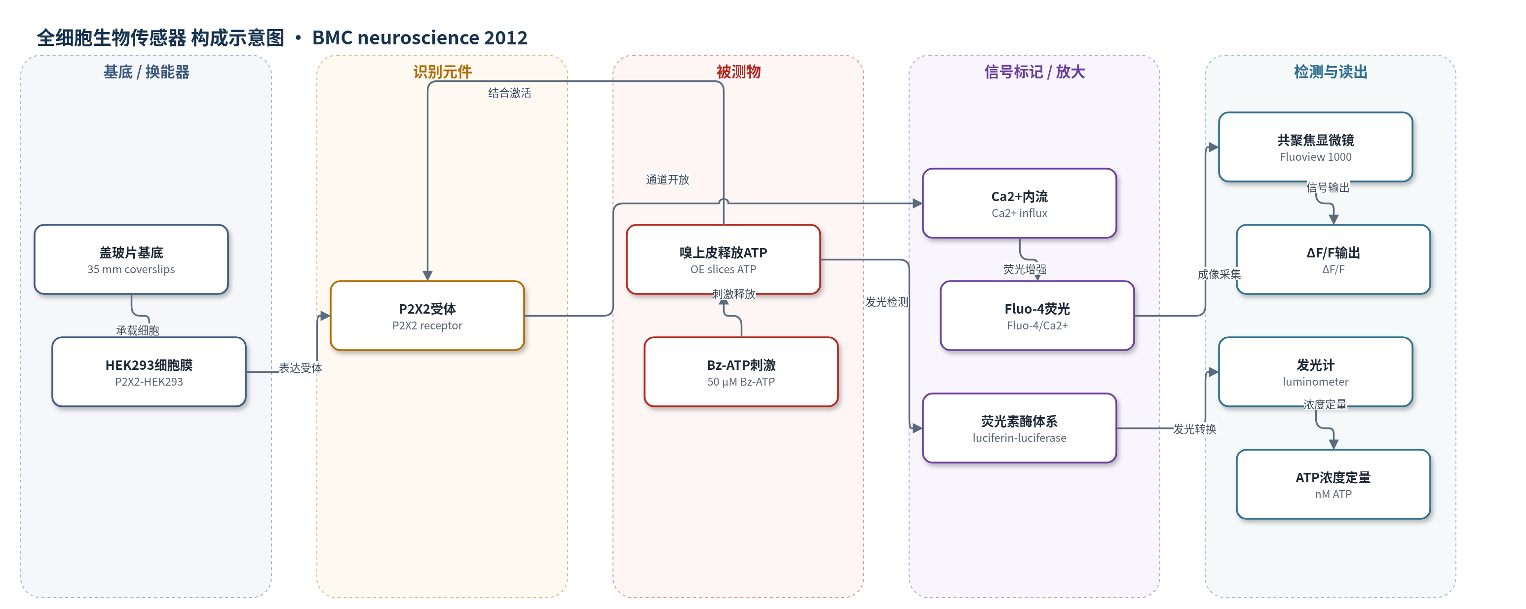
点击图片查看大图 · 依据论文自动绘制
传感器类型
全细胞生物传感器
检测对象
三磷酸腺苷(ATP);样品基质为小鼠新生嗅上皮(OE)切片在 Ringer 溶液中的释放液/共聚焦记录室。
检测原理
嗅上皮切片在 Bz-ATP 等嘌呤能激动剂刺激下,经钙依赖胞吐、P2X7/pannexin 通道或 ABC 转运体向培养液释放 ATP。释放的 ATP 扩散至 P2X2 转染 HEK293 全细胞生物传感器,激活膜上 P2X2 离子通道,引起 Ca2+ 内流;细胞内 Fluo-4 与 Ca2+ 结合后荧光增强,共聚焦显微镜记录 ΔF/F,其峰值随局部 ATP 浓度升高而增大。另一分支中,ATP 进入荧光素-荧光素酶体系,作为辅因子驱动荧光素氧化发光,发光强度经标准曲线换算为 ATP 浓度。该体系以受体-离子通道-钙指示剂实现识别与换能,并以酶催化发光提供定量读出。
检测灵敏度
线性范围: 10 nM - 5 μM;R^2 = 0.93–0.99
效应效果
全细胞生物传感器对 ATP 呈浓度依赖响应:50 nM、500 nM、1 μM 和 5 μM ATP 的峰值钙信号分别为 0.05±0.02、0.12±0.02、0.13±0.02 和 0.41±0.07 ΔF/F;PPADS 预处理将 5 μM ATP 响应降至 0.01±0.01 ΔF/F,表明响应依赖 P2X2。Bz-ATP 单独不激活 HEK293,但与嗅上皮切片共存时产生 0.34±0.04 ΔF/F,接近 5 μM ATP,提示切片释放约 5 μM ATP。荧光素酶法测得组成性释放约 19±4 nM,Bz-ATP 刺激组织释放 3108±914 nM,较无组织 576±424 nM 约增加 5 倍。作者认为该机制为嗅觉损伤后神经再生提供药靶。
传感器的构成
- 基底/培养载体:35 mm 盖玻片(coverslips),承载 P2X2 转染 HEK293 细胞并用于共聚焦成像。
- 识别/换能元件:P2X2 稳定转染 HEK293 细胞(P2X2-HEK293),表达 P2X2 嘌呤能受体,将 ATP 识别转换为 Ca2+ 内流。
- 信号标记物:Fluo-4 AM 钙荧光探针,进入细胞后螯合 Ca2+,Ca2+ 升高引起荧光增强。
- 样品源:Swiss Webster 小鼠新生嗅上皮切片(OE slices),在 Bz-ATP 刺激下释放 ATP。
- 读出系统:Olympus Fluoview 1000 共聚焦显微镜,采集 Fluo-4 的 ΔF/F 钙荧光信号。
中文摘要
本研究探讨新生小鼠嗅上皮中ATP的组成性释放与ATP诱导释放机制。采用荧光素-荧光素酶发光法定量ATP释放,并用共聚焦成像观察荧光ATP标记物quinacrine标记的囊泡ATP释放。结果显示,生理条件下ATP通过胞吐、半通道和ABC转运体组成性释放,可被艰难梭菌毒素A和丙磺舒抑制;ADP经P2Y受体和cAMP/PKA通路负向调节组成性释放。体内实验提示组成性ATP释放参与嗅上皮神经元稳态与增殖。ATP诱导的ATP释放可由ATP、αβMeATP、Bz-ATP和UTP等嘌呤能受体激动剂触发,并经钙依赖胞吐、激活的P2X1/7受体、P2X7-pannexin复合物或ABC转运体介导;PPADS、艰难梭菌毒素A、丙磺舒和羧苄西林可抑制该过程。P2X2转染HEK293生物传感器细胞钙成像进一步证实了诱导性ATP释放。
英文摘要
BACKGROUND: ATP is an extracellular signaling molecule with many ascribed functions in sensory systems, including the olfactory epithelium. The mechanism(s) by which ATP is released in the olfactory epithelium has not been investigated. Quantitative luciferin-luciferase assays were used to monitor ATP release, and confocal imaging of the fluorescent ATP marker quinacrine was used to monitor ATP release via exocytosis in Swiss Webster mouse neonatal olfactory epithelial slices.
RESULTS: Under control conditions, constitutive release of ATP occurs via exocytosis, hemichannels and ABC transporters and is inhibited by vesicular fusion inhibitor Clostridium difficile toxin A and hemichannel and ABC transporter inhibitor probenecid. Constitutive ATP release is negatively regulated by the ATP breakdown product ADP through activation of P2Y receptors, likely via the cAMP/PKA pathway. In vivo studies indicate that constitutive ATP may play a role in neuronal homeostasis as inhibition of exocytosis inhibited normal proliferation in the OE. ATP-evoked ATP release is also present in mouse neonatal OE, triggered by several ionotropic P2X purinergic receptor agonists (ATP, αβMeATP and Bz-ATP) and a G protein-coupled P2Y receptor agonist (UTP). Calcium imaging of P2X2-transfected HEK293 "biosensor" cells confirmed the presence of evoked ATP release. Following purinergic receptor stimulation, ATP is released via calcium-dependent exocytosis, activated P2X1,7 receptors, activated P2X7 receptors that form a complex with pannexin channels, or ABC transporters. The ATP-evoked ATP release is inhibited by the purinergic receptor inhibitor PPADS, Clostridium difficile toxin A and two inhibitors of pannexin channels: probenecid and carbenoxolone.
CONCLUSIONS: The constitutive release of ATP might be involved in normal cell turn-over or modulation of odorant sensitivity in physiological conditions. Given the growth-promoting effects of ATP, ATP-evoked ATP release following injury could lead to progenitor cell proliferation, differentiation and regeneration. Thus, understanding mechanisms of ATP release is of paramount importance to improve our knowledge about tissue homeostasis and post-injury neuroregeneration. It will lead to development of treatments to restore loss of smell and, when transposed to the central nervous system, improve recovery following central nervous system injury.