组成图示
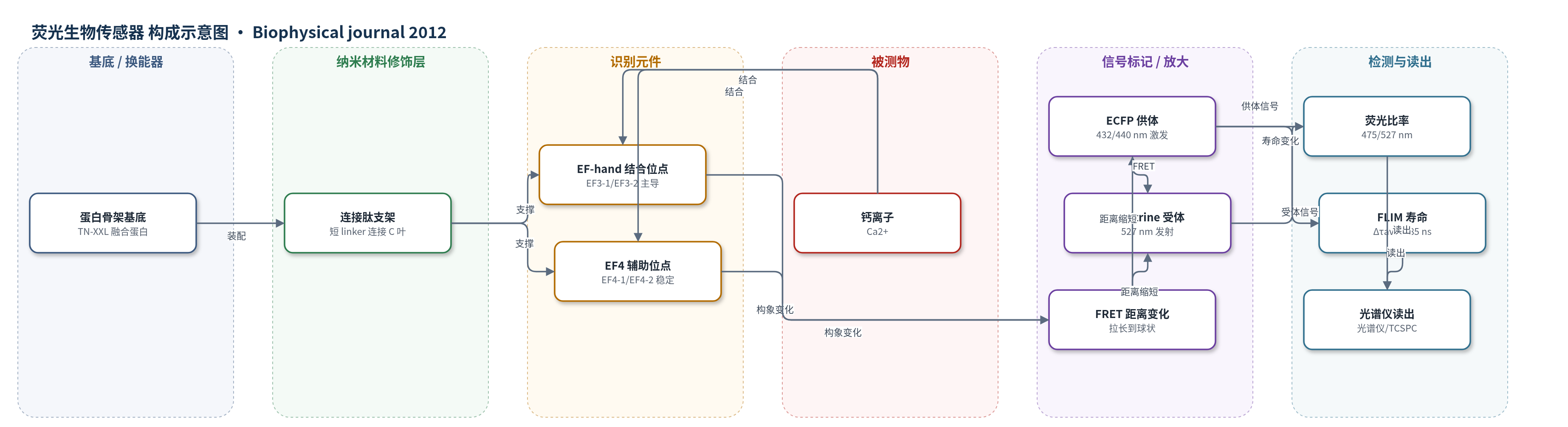
点击图片查看大图 · 依据论文自动绘制
传感器类型
荧光生物传感器
检测对象
钙离子(Ca2+);样品基质:体外缓冲液、细胞内/活体组织
检测原理
TN-XXL 是 ECFP–TnC C 端叶串联域–cpCitrine 融合蛋白。Ca2+ 结合到 TnC 的 EF-hand 位点,尤其 EF3-1 和 EF3-2,触发整个钙结合域发生协同构象变化:无钙时蛋白呈柔性拉长构象,ECFP 与 cpCitrine 相距较远,FRET 效率低;高钙时蛋白转变为刚性球状构象,两个荧光蛋白靠近,FRET 效率升高。该距离变化直接改变供体 ECFP 的荧光强度与寿命,并增强受体 cpCitrine 发射。检测可通过 475/527 nm 荧光比率或 FLIM 供体寿命读出,平均寿命变化 Δτave 约 0.85 ns。该传感器不依赖酶催化或核酸放大,信号由钙诱导构象变化直接换能。
检测灵敏度
原文未报告 LOD、线性范围、灵敏度斜率或 R^2;报告 Kd: 830 nM(FRET 强度法)、453 nM(荧光寿命法)
效应效果
TN-XXL 在高钙和无钙下均保持单体,无聚集;单荧光蛋白对照显示钙诱导构象变化不引起 ECFP 或 cpCitrine 的非 FRET 变化,高钙下无需假设非功能比例(受体质子化约 15%)。FRET 解离 τdecay 约 311 ms,与 EF3-1(317 ms)、EF3-2(277 ms)一致。温度升高使 Kd 从 830 nM 升至 1210 nM,τdecay 从 620 ms 降至 129 ms;pH 6.5 至 8.0 使 Kd 从 1030 nM 降至 451 nM,τdecay 从 620 ms 降至 425 ms。Δτave 0.85 ns 大于 YC3.6 的 0.46 ns,适合 FLIM。
传感器的构成
- 无外置基底:重组 TN-XXL 融合蛋白直接表达于细胞内,以蛋白自身构象变化为换能单元。
- 供体荧光蛋白:ECFP(enhanced cyan fluorescent protein),作为 FRET 供体,其发射/寿命随 Ca2+ 结合变化。
- 钙结合识别域:鸡骨骼肌肌钙蛋白 C(TnC)C 端叶串联重复(EF-hands 3/4,residues 94–162),特异性结合 Ca2+ 并触发构象变化。
- 受体荧光蛋白:cpCitrine(circularly permuted Citrine,YFP 衍生物),作为 FRET 受体,与 ECFP 距离变化改变 FRET 效率。
- 结构连接支架:短连接肽连接两个 TnC C 端叶,维持 EF-hand 排列并传递构象变化。
- 表达纯化标签:His 标签与 TEV 蛋白酶切割位点,用于 E. coli 表达、Ni-NTA 亲和纯化和去标签。
- 信号读出:荧光光谱/FLIM 系统,激发 ECFP(432/440 nm)并检测供体 475 nm、受体 527 nm 或寿命。
中文摘要
基因编码钙指示剂已成为在活体复杂组织和神经回路中成像信号的重要工具,但由于其人工多模块组成,结构-功能关系往往不清楚。本文结合蛋白质工程与动力学、光谱及生物物理分析,系统表征基于 Förster 共振能量转移(FRET)的钙生物传感器 TN-XXL。通过工程化酪氨酸荧光光谱,作者发现四个钙结合 EF-hand 中 EF3-1 和 EF3-2 主导 TN-XXL 的 FRET 输出,且这些位点的局部构象变化动力学与 FRET 变化一致。小角 X 射线散射和核磁共振表明,TN-XXL 在结合钙后由柔性拉长构象转变为刚性球状构象,从而产生 FRET 信号。研究还比较了荧光寿命光谱钙滴定与比率法,并评估可能影响荧光团的非 FRET 效应。这些数据详细刻画了 TN-XXL 的生物物理特性,可为 FRET 生物传感器的理性工程优化提供基础。
英文摘要
Genetically encoded calcium indicators have become instrumental in imaging signaling in complex tissues and neuronal circuits in vivo. Despite their importance, structure-function relationships of these sensors often remain largely uncharacterized due to their artificial and multimodular composition. Here, we describe a combination of protein engineering and kinetic, spectroscopic, and biophysical analysis of the Förster resonance energy transfer (FRET)-based calcium biosensor TN-XXL. Using fluorescence spectroscopy of engineered tyrosines, we show that two of the four calcium binding EF-hands dominate the FRET output of TN-XXL and that local conformational changes of these hands match the kinetics of FRET change. Using small-angle x-ray scattering and NMR spectroscopy, we show that TN-XXL changes from a flexible elongated to a rigid globular shape upon binding calcium, thus resulting in FRET signal output. Furthermore, we compare calcium titrations using fluorescence lifetime spectroscopy with the ratiometric approach and investigate potential non-FRET effects that may affect the fluorophores. Thus, our data characterize the biophysics of TN-XXL in detail and may form a basis for further rational engineering of FRET-based biosensors.