组成图示
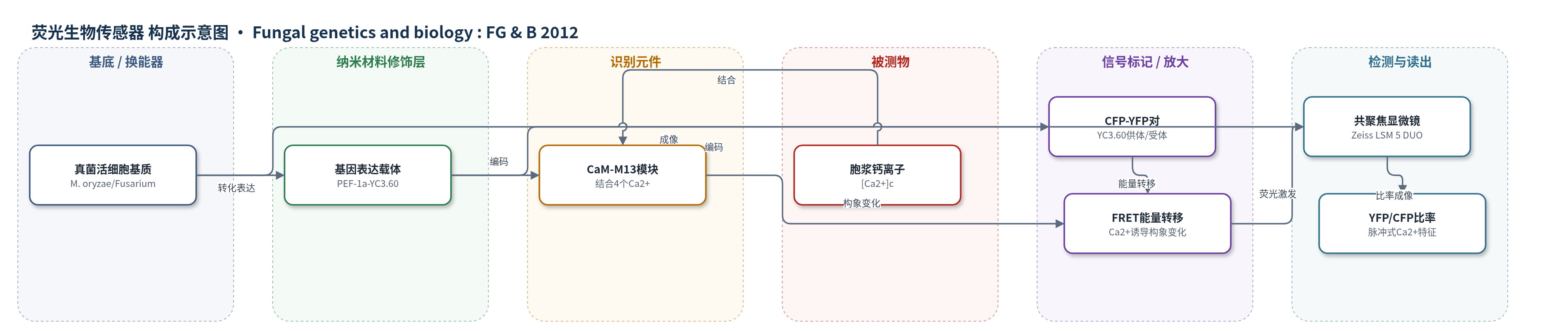
点击图片查看大图 · 依据论文自动绘制
传感器类型
荧光生物传感器
检测对象
胞浆游离钙离子(cytoplasmic free Ca2+, [Ca2+]c);样品基质:真菌活细胞(Magnaporthe oryzae、Fusarium oxysporum、Fusarium graminearum)及植株内组织(拟南芥根、水稻叶)
检测原理
YC3.60 由青色荧光蛋白(CFP)、钙调蛋白(CaM)、M13 钙结合模块和黄色荧光蛋白(YFP)串联组成,表达于真菌胞浆。当胞浆游离 Ca2+([Ca2+]c)升高时,CaM 结合 4 个 Ca2+ 并与 M13 相互作用,使 CFP 与 YFP 的相对距离和取向发生构象变化,从而增强 CFP 到 YFP 的 Förster 共振能量转移(FRET)。在 458 nm 激光激发下,CFP 发射减弱而 YFP 发射增强;通过分别采集 CFP 与 YFP 荧光并计算 YFP/CFP 比率,可比率化反映 [Ca2+]c 变化。比率成像可部分抵消表达量、光漂白和激发强度波动的影响,使传感器能够长时间、单细胞、亚细胞水平监测脉冲式 Ca2+ 信号。
检测灵敏度
原文未报告 LOD、线性范围、灵敏度斜率或相关系数。
效应效果
该传感器在三种真菌中稳定表达,可支持 30 min 至 1 h、间隔数秒的延时成像。三种物种呈现可区分 Ca2+ 特征:F. graminearum 生长速率 5.22 μm/min,脉冲平均 14.6 s(8.1–18.8 s);F. oxysporum 3.66 μm/min,平均 29.7 s(12.5–45.2 s);M. oryzae 3.05 μm/min,平均 22.4 s(12.4–30.0 s)。脉冲特征约 48 h 后更明显,并在菌丝分枝、隔膜形成、细胞接触、吸器形成和植株内生长中伴随大振幅瞬变。40 μM 4-bromo-A23187 引起持续 Ca2+ 内流并终止生长,100 μM 2-APB 诱发多次瞬变但生长继续。较染料和 aequorin,YC3.60 抗漂白、低自荧光,可单细胞成像,为真菌 Ca2+ 信号研究提供工具。
传感器的构成
- 样品基质/细胞载体:真菌活细胞(Magnaporthe oryzae、Fusarium oxysporum、Fusarium graminearum)及植物组织,提供胞浆 Ca2+ 微环境
- 表达载体:pBHt2-YC3.60 或 pBGt-YC3.60,含 EF-1a 启动子(PEF-1a)和 β-微管蛋白终止子(Tb-tubulin),驱动 YC3.60 表达
- 识别元件:钙调蛋白(CaM)与 M13 钙结合模块,结合 4 个 Ca2+ 后发生构象变化
- 荧光供体:青色荧光蛋白(CFP),458 nm 激发后发射青色荧光
- 荧光受体:黄色荧光蛋白(YFP),接受 FRET 能量后发射黄色荧光
- FRET 换能元件:CFP-YFP 对,Ca2+ 结合使 YFP/CFP 比率升高
- 光学读出:激光共聚焦显微镜(Zeiss LSM 5 DUO),采集 CFP/YFP 荧光并计算 YFP/CFP 比率
中文摘要
钙是通用信使,可将多种环境刺激和发育信号转化为特定细胞与发育反应。真菌虽普遍依赖 Ca2+ 信号调控生长、发育和致病,但缺乏可在单细胞水平成像亚细胞 Ca2+ 时空动态(即 Ca2+ 特征)的稳健方法,限制了相关研究。本文报道首次在真菌中成功表达基于 FRET 的 Ca2+ 生物传感器 Cameleon YC3.60。对表达该传感器的稻瘟菌 Magnaporthe oryzae、尖孢镰刀菌 Fusarium oxysporum 和禾谷镰刀菌 Fusarium graminearum 进行延时成像,结果显示胞浆 Ca2+([Ca2+]c)变化并非连续梯度,而是呈脉冲式波动,脉冲之间无明显梯度,且不同物种具有独特 Ca2+ 特征。脉冲式特征具有年龄和发育依赖性,并在菌丝分枝、隔膜形成、特化侵染结构分化、细胞间接触及植株内生长过程中出现显著 [Ca2+]c 瞬变。结合已测序基因组和真菌靶向基因操作手段,本文建立的数据、材料与方法将有助于解析 Ca2+ 介导的细胞与发育调控机制、极化生长形式以及真核生物中 Ca2+ 信号的演化。
英文摘要
Calcium is a universal messenger that translates diverse environmental stimuli and developmental cues into specific cellular and developmental responses. While individual fungal species have evolved complex and often unique biochemical and structural mechanisms to exploit specific ecological niches and to adjust growth and development in response to external stimuli, one universal feature to all is that Ca(2+)-mediated signaling is involved. The lack of a robust method for imaging spatial and temporal dynamics of subcellular Ca(2+) (i.e., "Ca(2+) signature"), readily available in the plant and animal systems, has severely limited studies on how this signaling pathway controls fungal growth, development, and pathogenesis. Here, we report the first successful expression of a FRET (Förster Resonance Energy Transfer)-based Ca(2+) biosensor in fungi. Time-lapse imaging of Magnaporthe oryzae, Fusarium oxysporum, and Fusarium graminearum expressing this sensor showed that instead of a continuous gradient, the cytoplasmic Ca(2+) ([Ca(2+)](c)) change occurred in a pulsatile manner with no discernable gradient between pulses, and each species exhibited a distinct Ca(2+) signature. Furthermore, occurrence of pulsatile Ca(2+) signatures was age and development dependent, and major [Ca(2+)](c) transients were observed during hyphal branching, septum formation, differentiation into specialized plant infection structures, cell-cell contact and in planta growth. In combination with the sequenced genomes and ease of targeted gene manipulation of these and many other fungal species, the data, materials and methods developed here will help understand the mechanism underpinning Ca(2+)-mediated control of cellular and developmental changes, its role in polarized growth forms and the evolution of Ca(2+) signaling across eukaryotic kingdoms.