组成图示
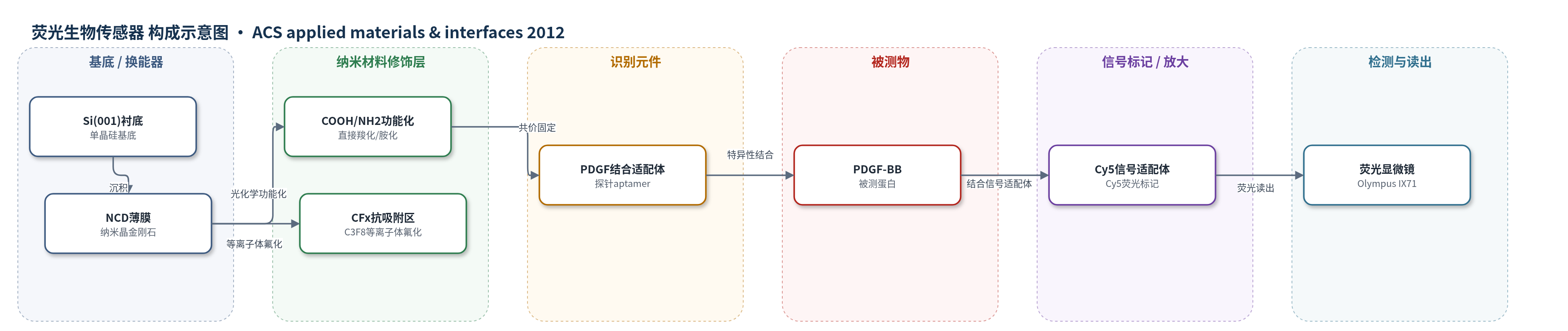
点击图片查看大图 · 依据论文自动绘制
传感器类型
荧光生物传感器
检测对象
血小板源性生长因子(PDGF,重点为PDGF-BB);样品基质:PBS缓冲液及含BSA的模拟血清蛋白基质(文中关联人血清/血浆)
检测原理
该传感器以直接羧化或胺化的纳米晶金刚石(NCD)表面为识别界面。点区内-COOH或-NH2经EDC/NHS活化形成NHS酯中间体,与5′端带COOH或NH2的PDGF结合适配体共价固定。当样品中的PDGF-BB进入界面时,被固定适配体特异性捕获;随后3′端标记Cy5的信号适配体与PDGF结合,使Cy5荧光染料富集于传感点区。荧光显微镜采集的Cy5荧光强度随PDGF-BB浓度增加而增强,在1000 nM至1 pM范围内呈浓度依赖响应。点区外C3F8等离子体氟化形成-CFx超疏水表面,抑制非特异吸附,提高信噪比。10% SDS洗涤可解离PDGF与Cy5适配体,实现再生。
检测灵敏度
LOD: 原文报告为“picomolar range”(未给精确数值);线性范围: 原文报告为“1 × 10^3 nM to 1 × 10^-3 nM”(1000 nM–1 pM);1 × 10^-2 nM仍显示较强荧光
效应效果
COOH-NCD与NH2-NCD传感器在PDGF检测中灵敏度和选择性相当。选择性测试中,ATP、葡萄糖氧化酶、脲酶和钙调蛋白在1000 nM下荧光信号极低;PDGF-AB信号约为PDGF-BB的一半,PDGF-AA几乎无信号。在含BSA的复杂蛋白基质中,固定PDGF浓度1000 nM时荧光强度无明显下降,表明抗血清蛋白干扰能力较强。经10% SDS再生后,两种传感器在1000 nM、1 nM和0.01 nM PDGF下均保持可分辨荧光,且一个月后仍无明显退化,显示长期稳定性和可重复使用性。作者认为其灵敏度高于此前基于金刚石晶体管的PDGF检测,具有临床应用潜力。
传感器的构成
- 基底衬底:Si(001)单晶硅,承载NCD薄膜。
- 传感基底:纳米晶金刚石(NCD)薄膜,MWPCVD沉积,提供稳定生物相容界面。
- 识别固定层:直接羧化(-COOH)或胺化(-NH2)NCD表面,作为适配体共价固定位点。
- 抗非特异吸附层:点区外C3F8等离子体氟化(-CFx)NCD表面,超疏水抑制非特异吸附。
- 活化偶联体系:EDC/NHS,形成NHS酯中间体,介导表面功能基团与适配体共价偶联。
- 识别元件:PDGF结合适配体(PDGF-binding aptamer),5′端NH2或COOH,特异性结合PDGF-BB。
- 信号标记物:Cy5标记信号适配体(Cy5-tagged aptamer),3′端Cy5荧光染料,结合PDGF后产生荧光。
- 再生试剂:10% SDS,洗涤去除PDGF与Cy5适配体,实现传感器再生。
中文摘要
本研究在纳米晶金刚石(NCD)薄膜表面设计了一种适配体传感器,用于检测血小板源性生长因子(PDGF)。通过直接光化学羧化和胺化,在NCD表面引入羧基(-COOH)和氨基(-NH2),作为固定PDGF结合适配体的探针单元。X射线光电子能谱(XPS)分析表明,直接胺化和羧化NCD表面的氨基与羧基覆盖率分别为23%和12%;尽管羧基密度较低,COOH-NCD生物传感器性能未受影响。荧光观察显示,COOH-NCD与NH2-NCD对PDGF蛋白检测具有相当的灵敏度和选择性。多次再生测试表明,两种传感器均保持高性能且无明显退化。
英文摘要
An aptasensor was designed on a nanocrystalline diamond (NCD) surface that combined with biological recognition elements, PDGF-binding aptamers, which inherently possess high affinity to PDGF-BB proteins. Functional components such as carboxylic acids (-COOH) and amines (-NH2) were directly introduced onto the NCD surface and used as probing units for immobilization of PDGF-binding aptamers. The surface coverage of different components on the NCD was analyzed by X-ray photoelectron spectroscopy (XPS) measurements, and the effects of various functionalizations on the NCD biosensor performance were investigated via fluorescence observations. The coverages of carboxyl and amine groups achieved were 12 and 23%, respectively, for the directly aminated and carboxylated NCD; however, the lower density of carboxyl groups on the functionalized surface did not deteriorate the performance of the COOH-NCD biosensor. Fluorescence investigations demonstrated comparable performance in sensitivity and selectivity for PDGF protein detection on COOH-NCD and NH2-NCD biosensors. Multiple regeneration tests clearly showed that the COOH-NCD biosensor as well as the NH2-NCD biosensor retained a high performance without exhibiting any noticeable degradation.