组成图示
点击图片查看大图 · 依据论文自动绘制
传感器类型
其他(动态光散射生物传感器)
检测对象
乙肝表面抗原(HBsAg);样品基质:Tris–HCl缓冲液、人血清(含HBV阳性/阴性、梅毒及HCV阳性血清)
检测原理
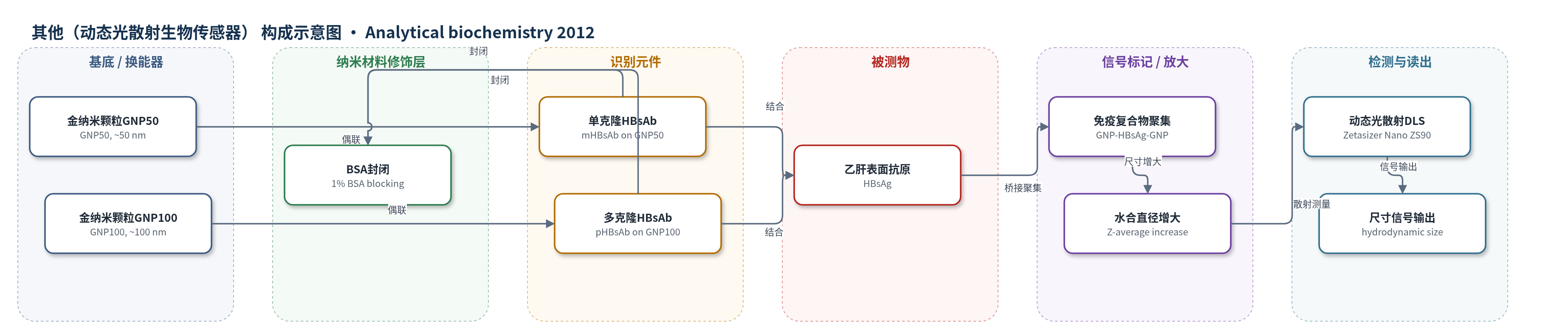
单克隆和多克隆乙肝表面抗体(HBsAb)分别物理吸附于GNP50和GNP100表面,形成两种纳米探针。加入HBsAg后,抗原作为桥通过抗原-抗体免疫反应同时结合两种抗体标记GNP,形成GNP-HBsAg-GNP聚集体;多克隆抗体还可引起GNP100自身聚集。免疫复合物使颗粒水合直径增大,布朗运动减慢,动态光散射(DLS)测得Z-average直径随HBsAg浓度增加而增大。由于GNP光散射截面大,双探针体系产生信号放大,无需洗涤和酶放大步骤,信号为尺寸变化而非颜色变化。
检测灵敏度
LOD: 0.005 IU/ml(GNP100–GNP50);LOD: 0.01 IU/ml(GNP10–GNP50);剂量依赖范围: 0.005–1 IU/ml;0.005 IU/ml (0.1 pM)
效应效果
该均相DLS纳米传感器在Tris–HCl缓冲液中检测HBsAg的LOD为0.005 IU/ml,比ELISA高80倍,比同一抗体对的局域SPR免疫传感器高2倍。在血清基质中可区分HBV阳性与阴性血清,并能排除梅毒和HCV阳性血清干扰;5个临床样本结果与ELISA一致。方法为一步、免洗、均相操作,探针制备简单,稳定性较好,作者认为可发展为自动化、广泛使用的生物传感器,并拓展至血浆、唾液、尿液等实际样品中其他蛋白标志物检测。
传感器的构成
- 纳米颗粒基底:金纳米颗粒(GNP,约10、50、100 nm),由氯金酸(HAuCl4)经柠檬酸钠还原制备,提供强光散射信号与抗体结合位点
- 识别元件:乙肝表面抗体(HBsAb),单克隆HBsAb偶联GNP50,多克隆HBsAb偶联GNP10/GNP100,通过物理吸附识别HBsAg
- 信号放大元件:GNP50与GNP100双探针组合,HBsAg桥接形成聚集体,增大水合直径并被DLS检测
- 封闭剂:含1 wt.%牛血清白蛋白(BSA)的Tris–HCl缓冲液(pH 7.4),洗涤封闭GNP剩余活性位点
- 检测介质:10 mM Tris–HCl缓冲液(pH 7.4)或人血清,用于抗原孵育与DLS测量
- 读出仪器:Zetasizer Nano ZS90动态光散射(DLS)系统,测量Z-average水合直径
中文摘要
本文构建了一种一步、免洗、均相纳米传感器检测方法,用于灵敏检测乙肝表面抗原(HBsAg)。该方法基于金纳米颗粒(GNP)的光散射性质,采用夹心免疫模式。研究将单克隆和多克隆乙肝表面抗体(HBsAb)分别偶联到不同直径的GNP上,形成两种纳米探针,并系统评估不同尺寸GNP组合的检测行为,确定最优组合。在Tris–HCl缓冲液中,由约50 nm和100 nm GNP组成的检测体系对HBsAg的检出限(LOD)为0.005 IU/ml,剂量依赖响应范围为0.005–1 IU/ml,表明其具有良好的诊断能力,可为低病毒载量蛋白标志物分析提供有用手段。透射电子显微镜(TEM)观察直接证明免疫反应诱导的聚集使颗粒水合直径增大。该检测在血清基质中分析HBsAg也表现出满意特异性。由于制备简单、读出方便且稳定性好,该方法有望发展为自动化、广泛使用的生物传感器检测。
英文摘要
In the current work, a one-step, washing-free, homogeneous nanosensor assay has been constructed to sensitively detect hepatitis B surface antigen (HBsAg) based on the light scattering property of gold nanoparticles (GNPs) through a sandwich model. The two nanoprobes in this study were designed by conjugating monoclonal and polyclonal hepatitis B surface antibody (HBsAb) onto the GNPs of different diameters. First, the detection behavior of the combinations of different sizes of GNPs was evaluated and the optimized combination was determined. In analyzing HBsAg in Tris-HCl buffer, such bioassay composed of GNPs of approximately 50 and 100 nm has a limit of detection (LOD) as high as 0.005 IU/ml and a dose-dependent response ranging from 0.005 to 1 IU/ml, which indicates its good diagnostic capability and provides a useful means to analyze protein biomarkers with low virus loads. Observation with transmission electron microscopy (TEM) provides direct evidence that the increase of hydrodynamic diameters resulted from the aggregation induced by immunological reactions. The bioassay also exhibits satisfactory specificity in analyzing HBsAg in serum media. Therefore, with its simple preparation, easy readout, and good stability, this bioassay has the potential to be developed into an automated and widely used biosensor assay.