组成图示
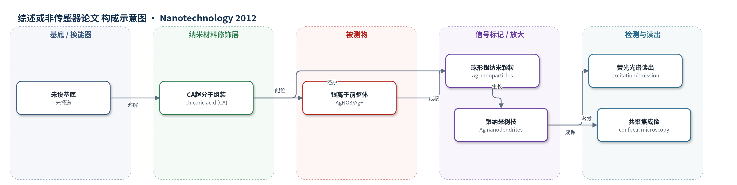
点击图片查看大图 · 依据论文自动绘制
传感器类型
综述或非传感器论文
检测对象
未报道(本文未构建传感器;样品为水相 AgNO3/CA 反应液)
检测原理
本文未建立针对特定分析物的传感检测原理,仅报道 CA 介导的 Ag+ 还原与定向生长机制。CA 的酚羟基和羧基先与 Ag+ 配位,随后发生氧化还原反应,将 Ag+ 还原为 Ag0 并形成球形 Ag 纳米颗粒。CA 分子通过氢键和配位形成超分子网络,控制银晶核的取向附着、晶面暴露和表面能,使纳米晶优先沿 [111] 和 [200] 方向生长,逐步形成主干、分支和叶状分层结构。随着纳米树枝尺寸增大和分支结构发展,局域表面等离子体共振(LSPR)高阶模式增强,激发峰由 410 nm 红移至 450、481 和 492 nm,发射峰由 560 nm 变为 570 nm,从而产生可见荧光。该发光来自 Ag 纳米树枝的等离子体相关光学响应,并非由被测物浓度驱动,因此未给出浓度-信号关系。
检测灵敏度
未报道(原文未给出 LOD、线性范围、灵敏度斜率或相关系数)
效应效果
论文未评估选择性、抗干扰、稳定性、重现性(RSD)或实际样品回收率,也未与 ELISA、HPLC、qPCR 对比。表征显示 CA 与 Ag+ 配位:FTIR 中 C=O 峰由 1724/1697 cm^-1 移至 1704/1690 cm^-1,OH 峰由 3474 cm^-1 移至 3465 cm^-1,出现 1557 cm^-1 醌式振动;HRTEM 晶格间距 0.204 nm,对应 fcc Ag {111}。在 CA 1 mM、AgNO3 0.5–5 mM 条件下,CA:Ag 约 1:2、Ag 1.5–2 mM 时最易形成树枝;3 周形成纳米树枝,4 周形成微蕨,3 个月形成超分支结构。激发峰由 410 nm 红移至 450、481、492 nm,发射峰由 560 nm 变为 570 nm,共聚焦显示发光。作者认为可用于光子学、成像、传感器或 SERS。
传感器的构成
- 基底/换能器:未报道(本文未构建传感器,仅在水相中合成纳米材料)
- 纳米材料修饰层:银纳米树枝(Ag nanodendrites),由硝酸银(AgNO3)在 chicoric acid(CA)存在下生物矿化形成
- 识别元件:未报道(CA 仅作为还原剂/导向剂,未作为抗体、适配体或 DNA 探针等识别元件)
- 信号标记物:未报道(Ag 纳米树枝自身发光,但未用于标记特定分析物)
- 封闭剂:未报道
- 电子供体:未报道
中文摘要
本文报道了在植物激素 chicoric acid(CA)存在下,通过温和条件下的单步生物矿化制备结构明确的银纳米树枝。CA 是一种已知的 HIV-1 整合酶抑制剂,在该体系中同时发挥还原剂和生长导向剂的双重作用:一方面其酚羟基和羧基可与 Ag+ 配位并参与电子转移,将 Ag+ 还原为 Ag0;另一方面 CA 形成的超分子网络可调控银晶核的取向附着与表面能,使纳米树枝主要沿 [111] 和 [200] 晶向生长。所得银纳米树枝具有高度有序的分层结构,并通过共聚焦显微镜观察到其发光特性。该研究展示了一种简单、环境友好的生物方法制备发光银纳米树枝,同时表明 CA 与银离子之间存在相互作用,可能为其在生物医学和生物传感器领域的应用提供线索。
英文摘要
In this work, we report the formation of well-defined silver nanodendrites via biomineralization under mild conditions in a single step, in the presence of the plant phytohormone chicoric acid (CA), a well-known HIV-I integrase inhibitor. CA played a dual role as reductant as well as directed the growth of the nanodendrites, which were found to grow primarily in the [111] and [200] directions. In addition to the formation of highly ordered hierarchical structures, the formed Ag nanodendrites were found to exhibit luminescence, as observed by confocal microscopy. This study not only demonstrates a new method for the preparation of luminescent silver nanodendrites using a simple, environmentally friendly biological method, but also indicates the ability of CA, a potent HIV-integrase inhibitor, to interact with silver ions which may shed light on its potential for additional biomedical and biosensor applications.