组成图示
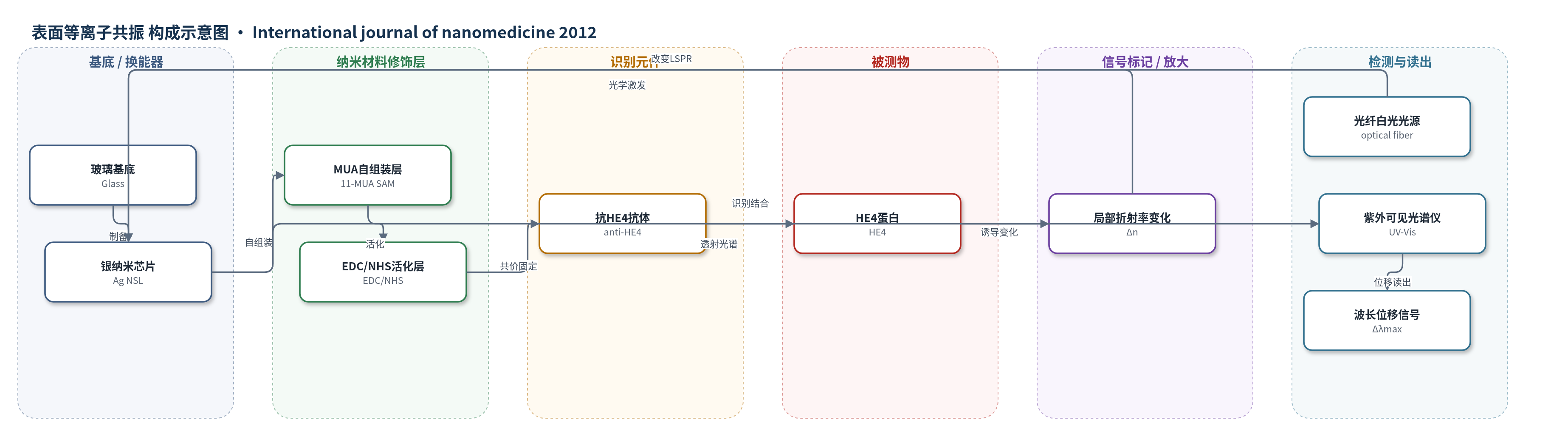
点击图片查看大图 · 依据论文自动绘制
传感器类型
表面等离子共振(SPR)生物传感器
检测对象
人附睾分泌蛋白4(HE4, human epididymis secretory protein 4),样品基质:人血清(human serum)及缓冲液标准品
检测原理
该传感器基于局部表面等离子共振(LSPR)对纳米颗粒附近局部折射率变化的敏感性。银纳米芯片表面经11-巯基十二烷酸自组装单分子层和EDC/NHS活化后,共价固定抗HE4抗体。当血清或缓冲液中的HE4与抗体特异性结合时,界面生物分子层厚度增加,导致银纳米颗粒周围局部折射率升高。根据Mie理论,LSPR消光峰位置随局部折射率变化而移动,因此HE4结合使消光峰向长波方向红移。HE4浓度越高,结合量越多,红移Δλmax越大。系统通过紫外-可见光谱仪实时记录消光峰波长,以Δλmax与HE4浓度对数建立定量关系,实现无标记、直接免疫检测。
检测灵敏度
LOD: 4 pM;线性范围: 10 pM–10,000 pM;线性回归: LSPR (nm) = 3.72 × log [HE4] (M) + 47.37;R = 0.997
效应效果
选择性:500 pM SCC和BSA无显著位移,无抗体芯片对HE4无响应。精密度:500 pM、5000 pM批内CV为9.29%、7.11%,批间CV为7.13%、7.17%。稳定性:8 M尿素再生4次CV 14.7%;4°C保存4周响应下降2.30%、7.21%、9.66%、17.09%,7次再生保留80%。临床:10例血清(5卵巢癌、5对照)LSPR与ELISA一致,卵巢癌红移11–17 nm,阴性<10.5 nm,浓度11.39–911.16 pM,相关系数0.926,P<0.05。检测40 min、无需预稀释,可作快速低成本无标记筛查替代。
传感器的构成
- 基底/换能器:玻璃基底(Glass)上制备银纳米芯片(Ag nanochip),采用纳米球光刻法(NSL)形成LSPR换能结构
- 自组装单分子层:1 mM 11-巯基十二烷酸(11-mercaptoundecanoic acid, MUA)在乙醇中孵育12 h,形成含羧基的SAM
- 活化层:75 mM EDC/15 mM N-羟基琥珀酰亚胺(NHS)活化MUA羧基,用于与抗体氨基形成酰胺键
- 识别元件:小鼠单克隆抗HE4抗体(anti-HE4, 10 µg/mL)共价固定,特异性识别HE4
- 封闭/失活层:1 M 乙醇胺(ethanolamine, pH 8.5)处理30 min,失活未反应酯基并降低非特异结合
- 样品结合层:HE4(human epididymis secretory protein 4)在缓冲液或人血清中与抗体孵育40 min
- 信号读出:紫外-可见分光光度计(UV-Vis spectroscope)配合CCD检测器记录LSPR消光峰波长位移(Δλmax)
中文摘要
人附睾分泌蛋白4(HE4)是卵巢癌早期诊断的重要生物标志物。本研究开发了一种基于局部表面等离子共振(LSPR)的无标记生物传感器,用于检测卵巢癌患者血清中的HE4。采用纳米球光刻法制备银纳米芯片,并以氨基偶联法将抗HE4单克隆抗体固定于芯片表面。检测时,HE4与固定抗体特异性结合,引起银纳米颗粒附近局部折射率变化,导致LSPR消光峰波长发生红移,通过紫外-可见光谱仪记录波长位移实现定量分析。该传感器检测速度快、特异性好、重现性佳且长期稳定。HE4线性范围为10 pM至10000 pM,检出限为4 pM。在人血清样品中,LSPR结果与酶联免疫吸附试验(ELISA)具有良好相关性。该研究首次将LSPR生物传感器应用于卵巢癌临床血清诊断,有望成为快速、低成本、无标记、便携的卵巢癌筛查工具。
英文摘要
BACKGROUND: Detection of the human epididymis secretory protein 4 (HE4) biomarker plays an important role in the early diagnosis of ovarian cancer. This study aimed to develop a novel localized surface plasmon resonance (LSPR) biosensor for detecting HE4 in blood samples from patients with ovarian cancer.
METHODS: Silver nanoparticles were fabricated using a nanosphere lithography method. The anti-HE4 antibody as a probe, which can distinctly recognize HE4, was assembled onto the nanochip surface using an amine coupling method. Detection was based on the shift in the extinction maximum of the LSPR spectrum before and after the HE4-anti-HE4 antibody reaction. These nanobiosensors were applied to detect HE4 in human serum samples and compare them using an enzyme-linked immunosorbent assay.
RESULTS: Tests relating to the detection of HE4 demonstrated that the LSPR-based biosensor featured a fast detection speed, good specificity, effective reproducibility, and long-term stability. The linear range for LSPR was between 10 pM and 10,000 pM, with a detection limit of 4 pM. An excellent correlation between LSPR and enzyme-linked immunosorbent assay results was observed in human serum.
CONCLUSION: This study is the first clinical diagnostic application of the LSPR biosensor in ovarian cancer. The LSPR biosensor, a rapid, low-cost, label-free and portable screening tool, can serve as a very effective alternative for the clinical serological diagnosis of ovarian cancer.